
Mastering Cell Division: Your Ultimate Guide to Mitosis and Meiosis with Effective Teaching Strategies for Biology Educators
Cell division is a fundamental process that keeps life going. It's how organisms grow, heal, and reproduce. Understanding mitosis and meiosis isn't just important for passing biology tests—it's essential for grasping how life continues from one generation to the next.
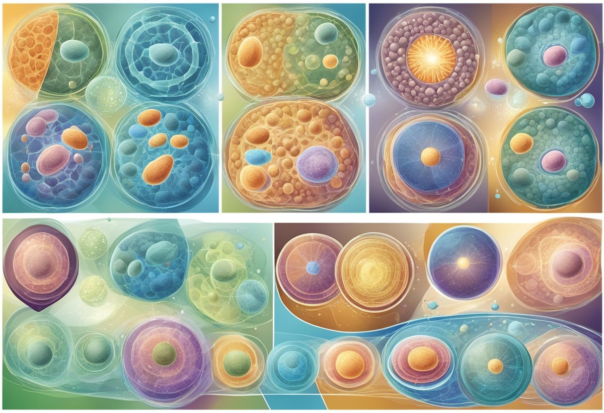
Teachers often struggle to make these complex concepts accessible to students. The microscopic world of chromosomes, spindle fibers, and cellular phases can seem abstract and disconnected from everyday life. This guide will bridge that gap with practical teaching strategies that make cell division engaging and comprehensible.
Whether you're a biology teacher looking for fresh approaches or a student trying to master these concepts, you'll find valuable techniques here. We'll explore visual models, hands-on activities, and digital tools that transform these invisible cellular processes into clear, memorable lessons.
Key Takeaways
Mitosis and meiosis serve different purposes in organisms, with mitosis creating identical cells for growth and repair while meiosis produces genetically diverse reproductive cells.
Effective teaching of cell division requires visual models, interactive activities, and real-world connections to make microscopic processes tangible for students.
Understanding the mechanisms of cell division provides insights into genetics, evolution, disease processes, and modern medical treatments like stem cell therapy.
Understanding Cell Division
Cell division powers life through carefully choreographed steps that ensure genetic material is properly distributed to new cells. This process maintains genetic continuity in both body cells and reproductive cells through different mechanisms.
Basics of Mitosis and Meiosis
Mitosis creates two genetically identical daughter cells with the same chromosome number as the parent cell. It occurs in somatic (body) cells and supports growth, tissue repair, and asexual reproduction.
Meiosis, by contrast, produces four genetically diverse cells with half the chromosomes of the parent cell. This reduction division happens only in specialized reproductive cells and enables sexual reproduction.
The key difference lies in their outcome: mitosis maintains chromosome number, while meiosis reduces it by half. This reduction is essential for preventing chromosome doubling when gametes combine during fertilization.
Both processes involve DNA replication followed by nuclear division, but meiosis includes two consecutive divisions without DNA replication between them.
The Cell Cycle and Its Phases
The cell cycle consists of interphase and the mitotic phase. Interphase includes three stages:
G1 phase: Cell growth and protein synthesis
S phase: DNA replication
G2 phase: Preparation for division
The mitotic phase includes:
Prophase: Chromosomes condense
Metaphase: Chromosomes align at cell's equator
Anaphase: Sister chromatids separate
Telophase: Nuclear envelopes form
Cytokinesis: Cytoplasm divides
Meiosis follows a similar pattern but undergoes two divisions—meiosis I and meiosis II. Meiosis I separates homologous chromosomes, while meiosis II separates sister chromatids, resulting in four haploid cells.
Regulatory Mechanisms
Cell division is tightly controlled by molecular checkpoints that monitor cellular conditions. These checkpoints ensure division occurs only when conditions are favorable.
The G1 checkpoint determines if a cell should enter the S phase or remain in G1. The G2 checkpoint confirms DNA has been properly replicated before mitosis begins.
Key regulators include:
Cyclins: Proteins that rise and fall in concentration during the cell cycle
Cyclin-dependent kinases (CDKs): Enzymes activated by cyclins that trigger specific cell cycle events
When these regulatory mechanisms fail, uncontrolled cell division may occur, potentially leading to cancer. External factors such as growth factors, nutrients, and cell density also influence when cells divide.
Cell size, DNA integrity, and chromosome attachment to spindle fibers are all monitored by checkpoint mechanisms to maintain genomic stability.
The Machinery of Mitosis
Mitosis is a precise cellular process that ensures accurate distribution of genetic material. The process involves distinct stages, specialized structures, and coordinated mechanisms that work together to maintain genetic stability.
Stages of Mitosis
Mitosis begins after the cell completes interphase, where DNA replication occurs. The first stage, prophase, involves chromosome condensation and nuclear membrane breakdown.
During prometaphase, spindle fibers attach to chromosomes at their centromeres. These fibers will guide chromosome movement.
Metaphase follows when chromosomes align at the cell's equator, forming the metaphase plate. This precise alignment is crucial for proper separation.
In anaphase, sister chromatids separate and move toward opposite poles of the cell. This movement is driven by spindle fibers that shorten and pull the chromatids apart.
Telophase marks the final stage where chromosomes reach the poles and begin to decondense. The nuclear membrane reforms around each set of chromosomes.
Chromosomes and Chromatids
Chromosomes are organized structures of DNA that become visible during cell division. Before mitosis, each chromosome replicates during interphase.
The replicated chromosome consists of two identical sister chromatids joined at the centromere. These chromatids contain identical genetic information.
Centromeres are specialized chromosome regions where spindle fibers attach. They play a critical role in ensuring proper chromosome movement during mitosis.
Kinetochores form at centromeres and serve as attachment sites for spindle fibers. This connection enables the precise movement of chromosomes during anaphase.
Condensins are proteins that help chromosomes condense into compact structures. This condensation is essential for preventing tangling during separation.
Cytokinesis and Its Importance
Cytokinesis follows mitosis and divides the cytoplasm to form two separate daughter cells. In animal cells, a contractile ring of actin filaments forms at the cell equator.
This ring contracts to create a cleavage furrow that deepens until the cell membrane pinches in two. The process ensures each new cell receives appropriate cellular components.
Plant cells use a different approach. They form a cell plate at the equator that grows outward to divide the cell. This plate eventually becomes the new cell wall.
Cytokinesis completes the cell division process by physically separating the cytoplasm. Without it, mitosis would create a single cell with multiple nuclei.
Proper cytokinesis ensures that each daughter cell receives one nucleus and approximately half of the cytoplasmic contents. This balance is essential for normal cell function.
Delving into Meiosis
Meiosis stands as the specialized cell division process essential for sexual reproduction, producing genetically diverse gametes with half the chromosomal count of the parent cell.
Distinction from Mitosis
Meiosis differs fundamentally from mitosis in both purpose and process. While mitosis creates identical daughter cells for growth and repair, meiosis produces gametes (eggs or sperm) with genetic diversity.
The key differences include:
Chromosome number: Mitosis maintains chromosome count; meiosis reduces it by half
Division cycles: One division in mitosis; two sequential divisions in meiosis
Genetic variation: Mitosis creates genetic copies; meiosis introduces variation through crossing over
These differences directly support reproduction by creating cells with haploid chromosome numbers that can combine during fertilization to form a complete set.
Meiosis I and II: A Closer Look
Meiosis occurs through two sequential divisions, each with distinct phases and purposes.
Meiosis I begins with DNA replication and homologous chromosome pairing. During prophase I, crossing over occurs where homologous chromosomes exchange genetic material, creating new gene combinations.
The homologous pairs separate in anaphase I, with each daughter cell receiving one chromosome from each pair. This reduction division is the key step that halves chromosome number.
Meiosis II resembles mitosis but without DNA replication beforehand. Sister chromatids separate, resulting in four haploid cells, each genetically unique.
This two-stage process ensures both chromosome reduction and genetic diversity essential for sexual reproduction.
Genetic Variability and Natural Selection
Meiosis creates genetic diversity through three key mechanisms:
Crossing over exchanges DNA segments between maternal and paternal chromosomes during prophase I, creating new gene combinations
Independent assortment of chromosomes during metaphase I allows random chromosome alignment
Random fertilization further increases variation when gametes combine
This genetic variability provides the raw material for natural selection. Beneficial mutations can spread through populations when they increase survival or reproductive success.
Genetic linkage (genes located close together on chromosomes) can influence how traits are inherited. Tightly linked genes tend to be inherited together, potentially preserving advantageous combinations.
This variability allows species to adapt to changing environments through natural selection, driving evolution over time.
Genetics and Inheritance
Genetics connects cell division to the larger picture of how traits pass from parents to offspring. The DNA in our chromosomes carries instructions that determine everything from eye color to disease risk.
From Genes to Proteins
Genes are segments of DNA that contain instructions for making proteins. This process begins with transcription, where DNA is copied into RNA in the nucleus. The RNA then travels to the cytoplasm for translation, where ribosomes read the genetic code and build proteins accordingly.
Proteins perform essential functions in the body, from enzymes that speed up chemical reactions to structural components that give cells their shape. The structure of proteins is critical to their function. Proteins begin as linear chains of amino acids that fold into complex three-dimensional shapes.
Mutations in DNA can alter the protein's structure or function. Some mutations have no effect, while others can lead to serious disorders by producing abnormal proteins or preventing normal protein production.
Patterns of Inheritance
Inheritance follows several predictable patterns first described by Gregor Mendel. These patterns explain how traits pass from generation to generation.
Common inheritance patterns include:
Dominant: Only one copy of the gene needed to express a trait
Recessive: Two copies of the gene needed to express a trait
Sex-linked: Genes located on sex chromosomes, often the X chromosome
Codominant: Both alleles are fully expressed
During meiosis, chromosomes shuffle and separate, creating unique combinations of genes in each reproductive cell. This genetic recombination explains why siblings look different despite having the same parents.
Some traits follow Mendelian inheritance patterns with clear dominant and recessive relationships. Others are polygenic, meaning multiple genes contribute to a single trait such as height or skin color.
The Role of Genetics in Cancer
Cancer develops when genetic mutations cause cells to divide uncontrollably. These mutations can affect three types of genes that regulate cell division.
Oncogenes promote cell growth when activated. Mutations can turn them on permanently, leading to excessive cell division. Tumor suppressor genes normally prevent excessive growth. When mutated, they lose their protective function.
DNA repair genes fix mistakes during DNA replication. When these genes malfunction, mutations accumulate more rapidly. This genetic instability accelerates cancer development.
Some cancer-causing mutations are inherited, creating predispositions to certain cancers. Most mutations occur during a person's lifetime due to environmental factors or random errors in DNA replication.
Modern cancer treatments increasingly target specific genetic mutations, allowing for more personalized and effective therapies.
Experimental Tools and Techniques
Scientists use specialized equipment and methods to study cell division processes. These tools allow researchers to observe chromosomes, analyze DNA, and perform controlled experiments on living cells.
Microscopy Essentials
Light microscopes help students view basic cell structures, but they lack the power to see detailed chromosomal movements. Compound microscopes with 400x-1000x magnification can reveal mitotic phases in onion root tips or whitefish blastula slides.
For advanced visualization, fluorescence microscopy uses special dyes that bind to DNA. This technique makes chromosomes glow against a dark background. Researchers often use DAPI stain, which emits blue light when attached to DNA.
Electron microscopes provide even greater detail, reaching magnifications of 10,000x or more. These powerful tools reveal intricate cellular structures involved in spindle formation and chromosome separation during both mitosis and meiosis.
Electrophoresis and Molecular Analysis
Gel electrophoresis separates DNA fragments by size, creating a visual "fingerprint" of genetic material. This technique helps scientists analyze chromosome content and detect abnormalities in cell division.
Common Electrophoresis Methods:
Agarose gel: Separates large DNA fragments
Polyacrylamide gel: Provides higher resolution for smaller fragments
Pulsed-field gel: Analyzes entire chromosomes
PCR (Polymerase Chain Reaction) amplifies specific DNA sequences, making them easier to study. Researchers use PCR to examine genes involved in cell cycle regulation and to detect mutations that disrupt normal division processes.
Fluorescence in situ hybridization (FISH) combines microscopy with molecular techniques. This method uses fluorescent probes to highlight specific chromosomal regions, helping identify abnormalities in chromosome number or structure.
Cell Culture and Experimental Design
Cell culture allows researchers to grow specific cell types in controlled laboratory conditions. Scientists commonly use HeLa cells (human cancer cells) to study mitosis because they divide rapidly and continuously.
Experimental design for cell division studies typically follows this process:
Form a testable hypothesis about a specific aspect of cell division
Design experiments with proper controls
Collect and analyze data
Accept or reject the null hypothesis based on statistical tests
Synchronization techniques help align cells at the same division stage. Common methods include serum starvation or chemical inhibitors that block specific cell cycle phases.
Modern studies often integrate multiple techniques. For example, researchers might use microscopy to observe dividing cells, molecular analysis to examine gene expression, and statistical tests to validate their findings in the primary literature.
Teaching Strategies for Cell Division
Effective teaching of cell division requires clear approaches that address common challenges students face. The right strategies can transform complex biological processes into accessible learning experiences.
Conceptual Framework and Misconceptions
Students often struggle with cell division concepts due to several common misconceptions. Many confuse mitosis and meiosis or believe that chromosomes and chromatids are the same thing. Teachers should begin by assessing prior knowledge through pre-tests or concept mapping exercises.
Visual models are essential tools for correcting these misconceptions. Three-dimensional models of chromosomes help students visualize the physical changes during different division phases. Comparison charts highlighting the differences between mitosis and meiosis can clarify their distinct purposes and outcomes.
It's important to connect cell division to everyday contexts. Discussing cancer as uncontrolled mitosis or explaining genetic diversity through meiosis makes abstract concepts more concrete for students.
Effective Tutorial Techniques
Hands-on activities significantly improve comprehension of cell division. Students can create chromosome models using pipe cleaners or colored clay to manipulate and demonstrate different division stages.
Proven tutorial approaches include:
Step-by-step animations that slow down complex processes
Cell division card sorts for sequencing practice
Microscope labs using prepared slides of dividing cells
Role-playing activities where students become chromosomes
Digital simulations allow students to manipulate variables and observe outcomes. These interactive tools help students visualize processes that are otherwise difficult to see.
Recording structured notes using graphic organizers helps students organize key concepts. Color-coding different stages or structures reinforces visual learning connections.
Supporting Student Understanding
Differentiated learning supports diverse student needs. Advanced students might explore chromosome abnormalities, while others benefit from additional review of basic concepts through simplified models.
Formative assessments should happen frequently throughout instruction. Quick checks like exit tickets or digital polls help identify confusion points before misconceptions become entrenched.
Effective support strategies:
Provide vocabulary guides with visual references
Create analogies relating cell division to familiar concepts
Offer guided practice before independent work
Use real-world examples relevant to student interests
Breaking complex processes into manageable chunks prevents cognitive overload. Teaching prophase before introducing metaphase builds conceptual understanding step by step.
Peer teaching opportunities help reinforce learning. When students explain concepts to classmates, they solidify their own understanding while providing alternative explanations.
Applications of Cell Division Knowledge
Understanding cell division opens doors to numerous practical applications in our world. The knowledge of mitosis and meiosis serves as a foundation for advancements in medicine, biotechnology, and our understanding of evolution.
Relevance in Medical Science
Cell division knowledge is crucial in diagnosing and treating diseases. Cancer develops when cells divide uncontrollably due to mutations in genes that regulate cell growth. Doctors use this understanding to develop targeted therapies that interrupt specific stages of mitosis in cancer cells.
Stem cell research relies on controlled cell division to regenerate damaged tissues. Scientists can now guide stem cells to differentiate into specific cell types for treating conditions like Parkinson's disease and spinal cord injuries.
Reproductive medicine also benefits from meiosis knowledge. Fertility specialists can identify chromosomal abnormalities that occur during meiosis, helping couples with conception difficulties understand their options.
Biotechnology and Genetic Engineering
Cell division principles enable scientists to manipulate genes and create new organisms. Techniques like CRISPR use the cell's natural division processes to edit DNA with unprecedented precision.
Agricultural applications include developing crops with improved traits:
Disease resistance
Higher yields
Better nutritional content
Drought tolerance
Pharmaceutical production has been revolutionized through cell division knowledge. Scientists can engineer bacteria and other organisms to produce insulin, vaccines, and other medications through controlled reproduction.
Cloning technologies depend on understanding how to reset a cell's division cycle, allowing scientists to create genetic copies of organisms for research.
Evolutionary Biology and the Tree of Life
Cell division mechanisms provide evidence for the relationships between species. The similarities in mitosis across different organisms support the theory of common ancestry.
Mutations that occur during DNA replication in cell division drive natural selection. These genetic changes create the variation that allows species to adapt to changing environments over time.
Scientists use differences in cell division processes to classify organisms on the tree of life. For example, the absence of a cell wall during division distinguishes animals from plants.
The study of meiosis helps explain the genetic diversity within populations. Through crossing over and independent assortment, meiosis creates unique combinations of genes that influence evolution's pace and direction.
Frequently Asked Questions
Cell division involves complex processes that often generate questions from both students and educators. These questions address the mechanics, differences, teaching strategies, and common misconceptions related to mitosis and meiosis.
What are the stages of mitosis and their functions?
Mitosis consists of five distinct stages: prophase, prometaphase, metaphase, anaphase, and telophase.
During prophase, chromatin condenses into chromosomes, and the nuclear envelope begins to break down. The nucleolus disappears, and spindle fibers start to form.
In prometaphase, the nuclear membrane completely dissolves. Kinetochores develop on the centromeres, and spindle fibers attach to these structures.
Metaphase involves the alignment of chromosomes at the cell's equator. This arrangement, called the metaphase plate, ensures proper chromosome distribution.
During anaphase, sister chromatids separate and move toward opposite poles of the cell. This separation occurs as the spindle fibers shorten.
Telophase marks the final stage where chromosomes reach the poles and begin to decondense. Nuclear membranes reform around each set of chromosomes, and the nucleoli reappear.
How do mitosis and meiosis differ and what are their respective roles in reproduction?
Mitosis produces two genetically identical daughter cells, while meiosis creates four genetically diverse cells. This fundamental difference reflects their distinct purposes.
Mitosis primarily functions in growth, development, and repair of somatic cells. It maintains the same chromosome number throughout generations of cells.
Meiosis serves reproductive purposes, creating gametes (eggs or sperm) with half the chromosomes of the parent cell. This reduction in chromosome number allows for genetic diversity when gametes combine during fertilization.
Meiosis includes two consecutive cell divisions with only one round of DNA replication. It also features crossing over, which further increases genetic variation.
What are the most effective strategies for teaching complex topics like cell division to students?
Visual aids significantly enhance understanding of cell division. Models, animations, and diagrams help students visualize the dynamic processes occurring during mitosis and meiosis.
Analogies and real-world comparisons make abstract concepts more relatable. Comparing chromosomes to recipe books or the spindle apparatus to puppet strings can help students grasp complex ideas.
Hands-on activities, such as modeling chromosomes with pipe cleaners or performing simulations, engage multiple learning styles. These activities transform passive learning into active participation.
Breaking down complex processes into smaller, manageable chunks prevents overwhelming students. Teaching one phase at a time allows for better comprehension before moving to the next concept.
Which cellular components are involved in the process of cell division, and what are their specific responsibilities?
Chromosomes carry genetic information and ensure its proper distribution to daughter cells. During division, they condense from loose chromatin to visible structures.
The centrosome serves as the main microtubule organizing center. It duplicates before division and forms the two poles of the mitotic spindle.
Spindle fibers, composed of microtubules, attach to chromosomes and facilitate their movement. They contract to pull chromosomes toward the cell poles.
The nuclear envelope temporarily disassembles during division to allow chromosome access to the spindle apparatus. It reforms around the separated chromosomes at telophase.
Centrioles, present in animal cells, help organize the spindle fibers. They replicate during interphase and migrate to opposite poles during prophase.
How can one distinguish between haploid and diploid cells during cell division?
Chromosome count provides the most definitive distinction between haploid and diploid cells. Diploid cells contain two complete sets of chromosomes (2n), while haploid cells have only one set (n).
Diploid cells typically undergo mitosis for growth and repair, maintaining their chromosome number. Haploid cells result from meiosis and function as gametes in sexual reproduction.
Cell size often differs between haploid and diploid cells, with haploid cells generally being smaller. This size difference reflects their reduced genetic content.
The presence of homologous pairs indicates a diploid cell. Haploid cells lack these pairs, containing only one chromosome from each pair found in the parent cell.
What common misconceptions do students have about mitosis and meiosis, and how can educators address them?
Students often confuse mitosis and meiosis, incorrectly using the terms interchangeably. Educators can address this by consistently using comparison charts and emphasizing their different functions.
Many students mistakenly believe that DNA replication occurs during division rather than interphase. Clear timelines showing when replication actually occurs can correct this misconception.
The concept of sister chromatids versus homologous chromosomes frequently causes confusion. Using different colored models helps students visually distinguish between these structures.
Students sometimes think that mitosis occurs only in plants and animals. Teachers should clarify that various forms of cell division occur in all eukaryotic organisms.
The idea that cytokinesis is part of telophase represents another common misconception. Educators should emphasize that cytokinesis is a separate process that often occurs alongside but distinct from mitosis.


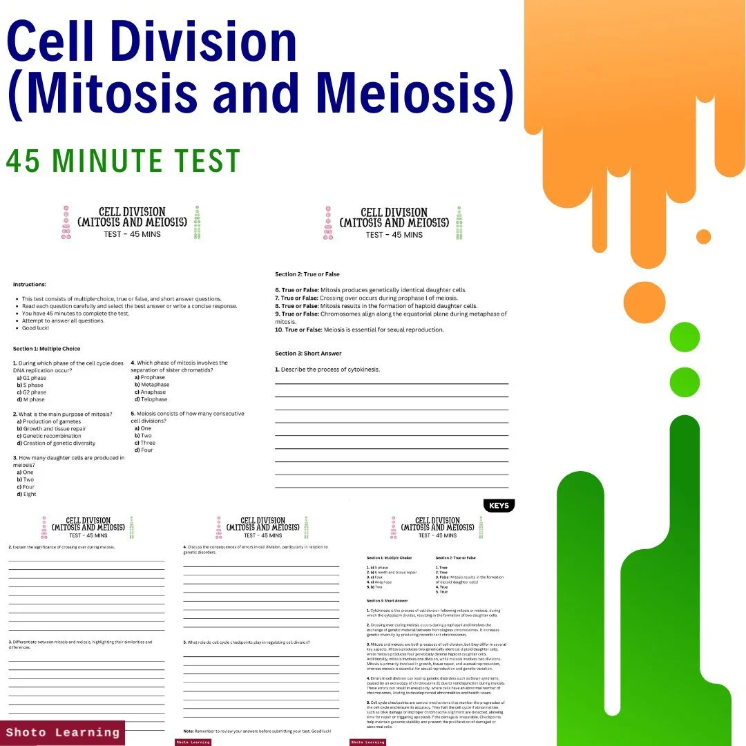


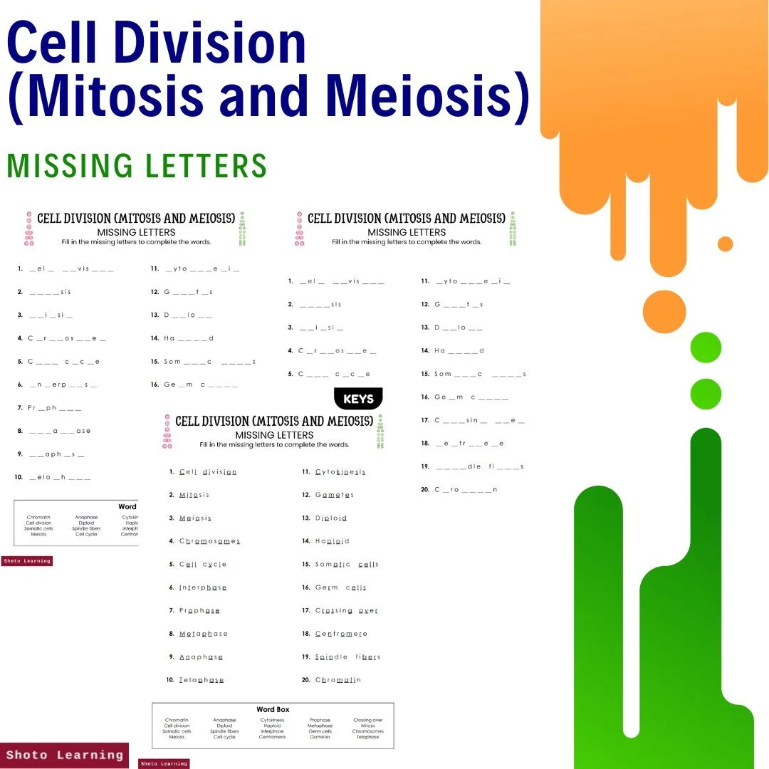


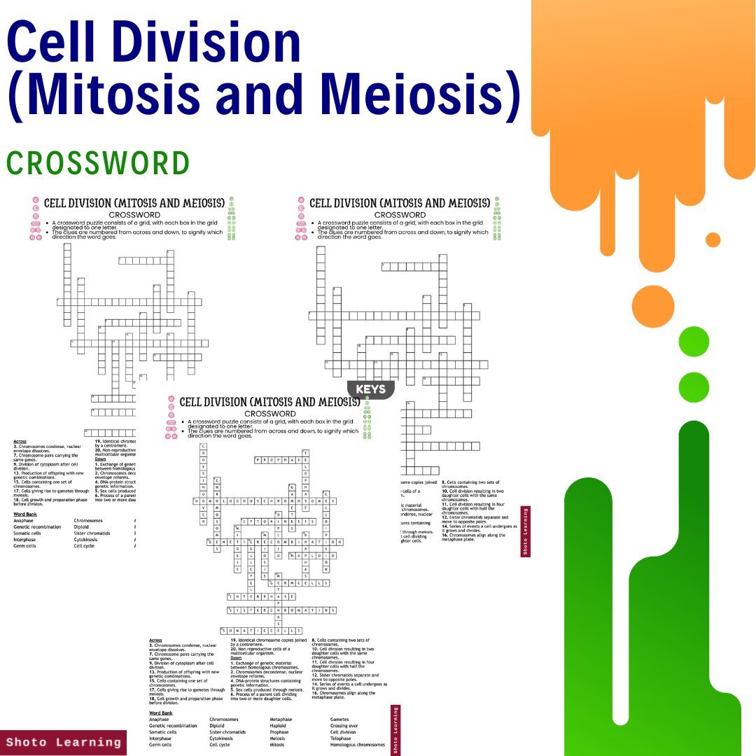


Unlock the mysteries of cellular reproduction and master the concepts of mitosis and meiosis with our Cell Division Mastery Bundle. This comprehensive collection of educational resources provides a wealth of engaging materials to enhance learning and understanding of cell division.
Key Features:
Assignments: Embark on 10 stimulating assignments covering diverse topics, encouraging critical thinking and practical application of knowledge.
Bingo - 30 Bingo Cards: Make learning fun with our interactive bingo game, featuring 30 cards filled with key terms and concepts related to cell division.
Crossword: Challenge your knowledge with a crossword puzzle that tests your understanding of mitosis and meiosis terminology.
Cryptogram: Decode secret messages related to cell division with our cryptogram activity, adding an element of mystery to your learning experience.
Debate: Engage in lively discussions and debates on cell division topics, fostering critical thinking and analytical skills.
Experiments: Explore hands-on experiments illustrating fundamental principles across various disciplines, fostering deeper understanding and engagement.
Fill in the Blank: Reinforce your understanding of cell division concepts with fill-in-the-blank exercises that provide practice and review.
Flashcards: Study on the go with our flashcards, featuring essential terms and definitions related to mitosis and meiosis.
Free ABC Order: Practice organizing key terms alphabetically with our free ABC Order worksheet, reinforcing vocabulary skills.
Free Story Book: Dive into the world of cellular reproduction with our free storybook, offering an engaging narrative on the processes of mitosis and meiosis.
Free Poster: Decorate your learning space with our free poster, providing a visual overview of cell division processes and stages.
Lesson Plan: Dive deep with our detailed lesson plan, complete with engaging activities and discussion topics.
Matching: Test your knowledge of cell division terminology with our matching activity, pairing terms with their corresponding definitions.
Missing Letters: Challenge yourself with our missing letters worksheet, filling in the blanks to complete cell division-related words.
Multiple Choice: Assess your understanding of cell division concepts with multiple-choice questions that cover a range of topics.
PowerPoint: Explore the intricacies of mitosis and meiosis with our informative PowerPoint presentation, featuring detailed explanations and visual aids.
Questions and Answers: Test your knowledge with a set of questions and answers that cover key concepts in cell division.
Reading Comprehension: Sharpen reading skills with engaging passages, perfect for students and educators alike.
Scramble: Unscramble cell division-related words with our scramble worksheet, sharpening your vocabulary and spelling skills.
Test (45 Minutes): Evaluate your understanding of mitosis and meiosis with a 45-minute test that covers essential topics in cellular reproduction.
True or False: Determine the accuracy of statements related to cell division with our true or false activity.
Word Search: Have fun while reinforcing vocabulary with our word search puzzle, featuring cell division-related terms hidden within.
Writing Prompts: Stimulate critical thinking and creativity with a set of writing prompts that explore various aspects of cell division.
Embark on a journey of discovery and mastery with our Cell Division Mastery Bundle. Whether you're a student, educator, or lifelong learner, these engaging resources provide valuable tools and insights to deepen your understanding of mitosis and meiosis.
Pages: 135 pages
Order your copy today!