
How to Teach Cell Division: Effective Strategies for Simplifying Complex Biological Processes
Cell division is one of the most important processes for all living things, but it can be tough for students to understand. Teachers can make cell division easier to learn by breaking down each step and using simple visuals. With the right approach, any student can follow these ideas and feel confident about why cells divide and how it happens.
A lesson on cell division becomes much clearer with hands-on activities, clear models, and easy-to-understand terms. When students can see and touch what they learn, concepts like mitosis and meiosis feel less overwhelming and more logical.
Key Takeaways
Explaining cell division with simple steps helps students understand.
Interactive tools and hands-on activities make learning easier.
Addressing myths and common mistakes improves comprehension.
Understanding Cell Division
Cell division is a core idea in cell biology. It helps explain how living things grow, repair, and reproduce. The cell cycle controls the steps cells take as they get ready to divide and make new cells.
Purpose and Importance
Cell division lets organisms grow, develop, and heal. For multicellular organisms, this means damaged tissue can be replaced or older cells can be renewed. For example, skin heals after a cut because new cells are made through cell division.
One main purpose is to produce two identical daughter cells from one parent cell. This helps keep genetic information stable across generations. Without cell division, growth and repair would not happen.
Cell division can also lead to mutations, which are changes in DNA. While some mutations are harmful, others drive evolution by introducing new traits. This process shapes the diversity of life over time.
Overview of Cell Cycle
The cell cycle is the series of steps that cells follow as they grow and divide. There are two main types of cell division: mitosis and meiosis. Mitosis happens in most cells for growth and repair, while meiosis makes gametes like sperm and eggs.
The cell cycle has different stages:
G1 phase (growth)
S phase (DNA synthesis)
G2 phase (growth and preparation)
M phase (mitosis or meiosis and division)
Cells use checkpoints to make sure each stage is complete before moving on. This helps prevent mistakes. If the process goes wrong, it can lead to problems, such as cancer, which is uncontrolled cell growth.
Exploring Mitosis
Mitosis is a process that allows cells to make identical copies of themselves. During mitosis, cells carefully move and split their chromosomes so that each new cell (called a daughter cell) gets a full set.
Phases of Mitosis
Mitosis has several steps, or phases: prophase, metaphase, anaphase, and telophase. These happen after the S phase, when chromosomes are copied in preparation for cell division.
Prophase: Chromosomes condense and can be seen with a microscope. The nuclear membrane starts to break down.
Metaphase: Chromosomes line up in the middle of the cell. This position is called the metaphase plate.
Anaphase: Each chromosome splits into two. Sister chromatids are pulled to opposite sides of the cell.
Telophase: New nuclear membranes form around each set of chromosomes, and the cell is almost ready to divide.
After these phases, cytokinesis splits the cell into two daughter cells, each with the same number of chromosomes as the original cell.
Chromosome Condensation and Separation
At the start of mitosis, chromosomes go through condensation. This process packs chromosomes into tight, visible shapes, making them easier to move and less likely to get tangled.
During anaphase, separation begins. The sister chromatids (identical halves of a copied chromosome) separate and move to opposite sides of the cell. This makes sure each daughter cell gets a complete set of chromosomes.
Errors in condensation or separation can cause cells to get the wrong number of chromosomes. This can lead to problems for the new cells, making careful control of these steps very important.
Role of Spindle Formation
Spindle fibers are special structures made of proteins. They form during prophase and are essential for moving chromosomes.
The spindle comes from areas called centrosomes. Spindle fibers attach to chromosomes at a region called the centromere. Once attached, these fibers pull the chromosomes apart during anaphase.
Having a strong and well-formed spindle is necessary for correct chromosome movement. Without proper spindle formation, chromosomes might not separate evenly, causing mistakes in cell division.
Introducing Meiosis
Meiosis is a type of cell division that reduces the chromosome number by half, creating gametes for sexual reproduction. This process leads to genetic diversity among the offspring of species because it mixes genes in unique ways.
Stages of Meiosis
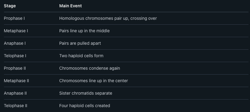
Meiosis takes place in two main stages: Meiosis I and Meiosis II. Each stage divides cell contents differently. In Meiosis I, homologous chromosomes pair up and then separate into different cells. This reduces the chromosome number from diploid (two sets) to haploid (one set).
Meiosis II is similar to mitosis. Here, the sister chromatids of each chromosome split apart. At the end of both stages, four new cells are made. Each has half the original number of chromosomes.
A summary of meiosis stages:
Differences Between Mitosis and Meiosis
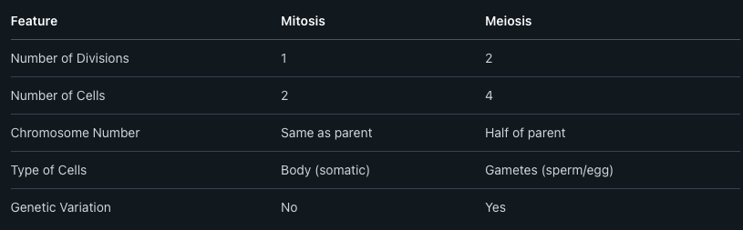
Mitosis and meiosis are both forms of cell division, but they serve different roles. Mitosis results in two cells that are genetically the same as the parent cell. These cells keep the chromosome number the same and are used for growth and repair.
Meiosis happens only in cells that form gametes, such as eggs and sperm. It creates four cells, each with half the chromosome number of the original. Meiosis also shuffles the DNA to create genetic differences.
Comparison Table:
Genetic Diversity and Its Significance
Meiosis increases genetic diversity, which is important for the survival of species. During meiosis, homologous chromosomes exchange genetic material in a process called crossing over. This mixes genes in new ways.
Random assortment also occurs when chromosomes are separated into gametes. Each gamete has a different mix of genes. When gametes from two parents combine during sexual reproduction, offspring inherit new combinations of traits.
This genetic variety helps populations adapt to changing environments. It lowers the chance that all individuals will be affected by the same disease or problem. Because of meiosis, each new generation has combinations of traits different from their parents.
Key Cellular Components
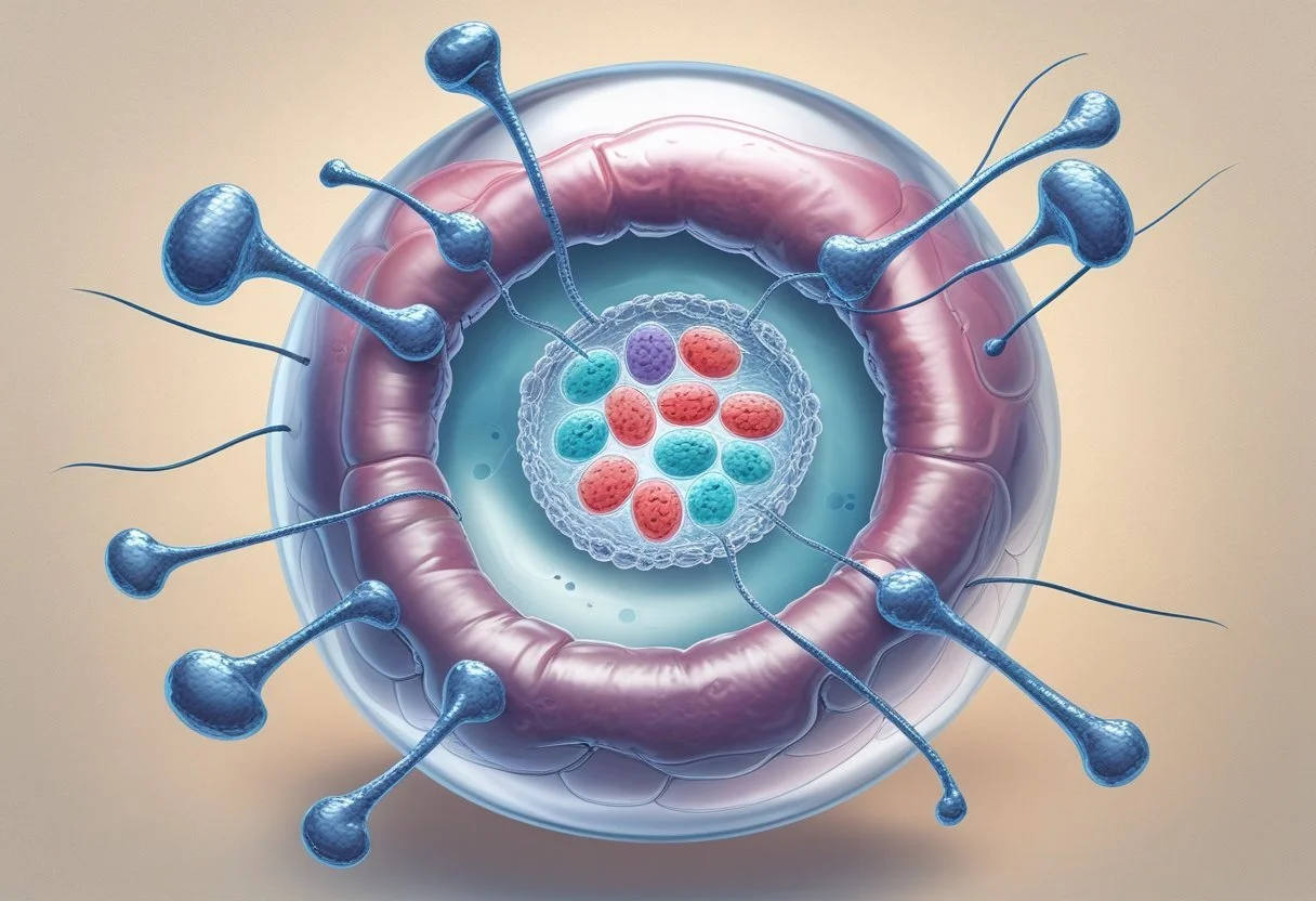
Cell division depends on specific parts inside the cell. DNA, proteins, the cell nucleus, and organelles all play special roles in how cells copy themselves and share genetic information.
Role of DNA and Genetic Material
DNA carries all the genetic information in a cell. It is shaped like a twisted ladder, called a double helix. Each DNA molecule is made of smaller units called nucleotides.
Before a cell divides, it must copy its DNA during a process called replication. This ensures that each new cell gets a complete set of instructions. DNA contains genes, which tell cells how to make proteins and control their activities.
Key points:
DNA is the blueprint for building and running the cell.
Errors in copying DNA can lead to mutations.
The cell checks and repairs its DNA during division to keep genetic material accurate.
Proteins and Organelles in Cell Division
Proteins help control every step during cell division. Some proteins copy DNA, while others guide and pull apart chromosomes.
Organelles support cell division by providing energy and organizing the contents of the cell. The centrosome is important for building the spindle fibers that separate chromosomes. The mitochondria supply energy so the process can happen.
Main proteins and organelles involved:
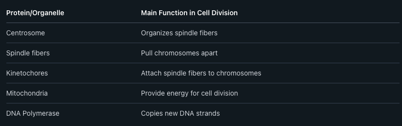
Proteins and organelles work together to keep division orderly and accurate.
Cell Nucleus and Chromosome Structure
The cell nucleus is a membrane-bound area that holds DNA. During cell division, the nucleus keeps genetic material secure until it is time to share it between cells.
Chromosomes are long strands of DNA packaged tightly with proteins. In human cells, there are 46 chromosomes that are copied before division. Chromosome structure allows the DNA to be moved safely without breaking.
Nuclear membranes disappear when cell division begins. Chromosomes line up, then each copy moves to a new cell. This organized structure prevents loss or damage to genetic material. Cells depend on the nucleus and chromosomes to manage genetic information through each division step.
Asexual Reproduction Mechanisms
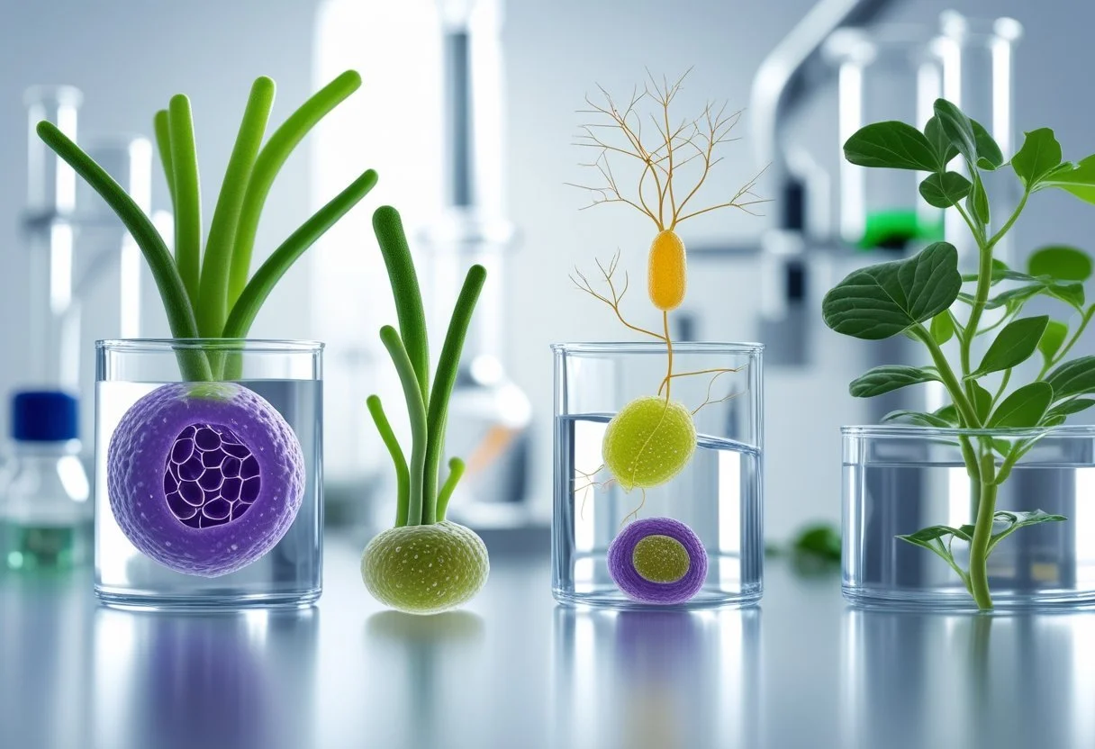
Asexual reproduction allows organisms to make offspring without a partner. Cells divide and create new cells, copying their genetic material so each new cell has the same chromosome number as the original.
Binary Fission
Binary fission is the most common method of asexual reproduction in single-celled organisms like bacteria.
During binary fission, the cell first copies its single circular chromosome. The chromosome attaches to the cell membrane. As the cell grows, it stretches and the two chromosomes move apart. The cell then splits, dividing the cytoplasm evenly and creating two identical daughter cells.
Each new cell gets a full set of DNA. In the table below, compare key features:
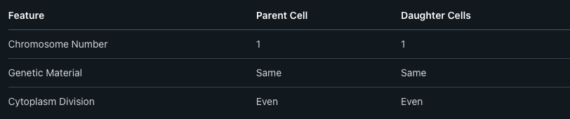
This process is quick and can happen in as little as 20 minutes under the right conditions. It helps bacteria adapt and multiply rapidly.
Other Asexual Processes
There are other ways organisms reproduce asexually besides binary fission.
Budding is one method. Yeast cells, for example, grow a small bulge or "bud" out of the parent cell. The bud gets a copy of the chromosome and some cytoplasm, then breaks away to become a new cell.
Fragmentation occurs in some simple animals and plants. The parent breaks into pieces, and each piece can grow into a new individual with the same chromosome number.
Vegetative reproduction in plants uses structures like runners or tubers. Each new part forms a complete plant that is genetically identical to the parent plant.
Asexual reproduction ensures that offspring are genetically identical to their parents. This can be useful in stable environments, as all new organisms get traits that already work well.
Sexual Reproduction and Its Implications
Sexual reproduction mixes genetic material from two parents, creating offspring that are different from each parent. This process relies on special cells called gametes and influences genetic diversity in a population.
Formation of Gametes
Gametes are reproductive cells. In animals, these are egg cells in females and sperm cells in males. Plants also have gametes, usually called pollen and ovules.
Formation of gametes happens through a process called meiosis. Meiosis is different from mitosis because it reduces the number of chromosomes by half. For humans, normal body cells have 46 chromosomes, but gametes have only 23.
Meiosis shuffles genetic material. It randomly splits and recombines chromosomes, which leads to each gamete being unique. This is why siblings can look different even though they have the same parents.

Genetic Variation in Offspring
When gametes from two parents join, the offspring gets half of its DNA from each parent. This combination creates new gene mixtures. These new mixtures cause differences in hair color, eye color, height, and many other traits.
Genetic diversity is important because it helps populations survive changes in the environment. If every individual were the same, a disease could wipe out all of them. With diversity, some might survive and pass on helpful traits.
Mechanisms that increase variation include:
Crossing over during meiosis
Random sorting of chromosomes
Fertilization combining two unique gametes
Genetic variation is a key reason why no two people (except identical twins) are exactly alike.
Zygote Development
After fertilization, the gametes fuse to form a zygote. The zygote now has a complete set of chromosomes, with half from each parent.
The zygote starts dividing right away. It grows through mitosis, forming all the cells in the new organism. In humans, this leads to the development from a single cell to a fully formed baby.
Stages of zygote development:
Fertilization (formation of zygote)
Mitosis (cell division)
Specialized cell formation (differentiation)
Each stage is important for the healthy growth of the offspring. Proper development results in genetically unique individuals, continuing the cycle of life.
Effective Teaching Strategies for Cell Division
Breaking cell division into smaller steps helps students understand each part of the process. Using visuals and hands-on activities can make tough ideas easier to grasp for learners with different needs.
Simplifying Complex Processes
To explain cell division, teachers can first introduce the main phases of mitosis and meiosis in order: prophase, metaphase, anaphase, and telophase. Using a step-by-step approach lets students build knowledge bit by bit.
Simple charts and diagrams can show how chromosomes line up and separate. Teachers can give examples from real life, such as how cells heal a cut or help plants grow.
Breaking the process into a list or flowchart helps students remember the main points:
Prophase: Chromosomes appear.
Metaphase: Chromosomes line up in the middle.
Anaphase: Chromosomes move apart.
Telophase: New nuclei form.
Quizzes and short writing activities can help students explain each step in their own words.
Visual and Interactive Tools
Using models, slides, and digital animations allows students to explore the stages of cell division more closely. Hands-on activities, like using yarn or colored beads to model chromosomes, can help students see what is happening.
A table that shows each stage and key events can make differences between mitosis and meiosis clearer:
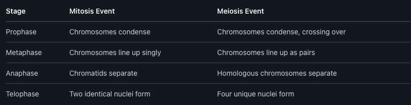
Teachers may extend lessons by giving students models to build or digital interactives to complete. Group work can let students share what they explored and learned.
Tailoring Approaches to Student Needs
Students have different learning styles, so teachers should adjust their approaches. For visual learners, diagrams and drawings are important. Others may benefit more from talking through the process or acting it out.
Some students need extra support. Teachers can give guided notes, step-by-step explanations, or extra time to explore diagrams. Advanced learners may extend their studies by comparing cell division in animals and plants.
Flexible activities, such as small group projects or review games, help address various needs. Using clear language and checking for understanding ensures all students can follow and explain cell division well.
Hands-On Classroom Activities
Practical activities help students see how cell division works in real time. Using models, acting out the phases, and applying technology can build a stronger understanding of chromosome movement and the changes cells go through.
Modeling Chromosome Movement
Teachers can use items like colored yarn, pipe cleaners, or beads to represent chromosomes. In groups, students show how chromosomes line up during metaphase, split during anaphase, and move to new cells in telophase.
A table can help students track chromosome positions:
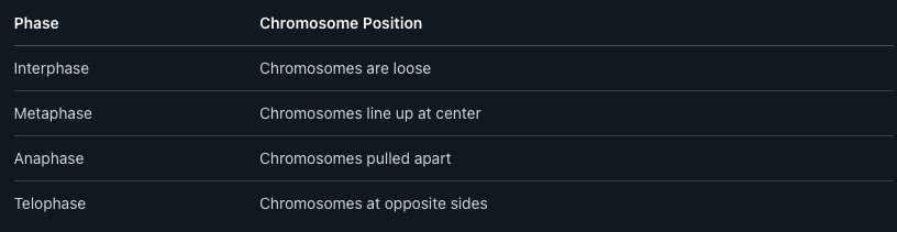
Building these models makes abstract ideas concrete. Students can touch and move "chromosomes," which supports memory and understanding.
Role-Playing the Phases
Role-playing turns students into cell parts. Each student can be a chromosome, spindle fiber, or cell membrane. In this activity, the process of mitosis is broken down into steps.
During interphase, "chromosomes" loosely move around. In metaphase, they line up in the middle, and during anaphase, they are pulled to opposite ends. Telophase ends the role-play as new "cells" form.
Teachers give clear instructions to keep each round focused. This method helps students remember what happens at each phase of cell division.
Utilizing Technology and Simulations
Computer simulations and interactive apps let students see cell division at a microscopic level. Programs such as CellCraft or online simulations let users control or view chromosome movement across all phases.
Animations illustrate how chromosomes double, line up, split, and move apart. Some tools allow color-coding for each stage, making metaphase, anaphase, and telophase easy to spot.
This technology helps when microscopes aren't available, providing a close look at cell behavior and a repeatable way to review each phase. Students can pause, rewind, and focus on key changes in chromosomes as cells divide.
Assessment and Evaluation Methods
Strong assessment strategies help teachers understand how well students grasp the steps and parts of cell division. Using clear methods allows teachers to check for understanding, adjust lessons, and support all learners.
Formative Assessment Techniques
Formative assessment tools give instant feedback during lessons. Teachers can use quick quizzes, diagrams, or worksheets to see if students understand mitosis and meiosis steps. Exit tickets are helpful for checking key points at the end of class.
Peer teaching is another effective technique. Students can work in pairs to explain a process like chromosome separation using drawings or short talks. This helps the teacher spot misunderstandings.
Teachers may create checklists for each stage of cell division. For example:
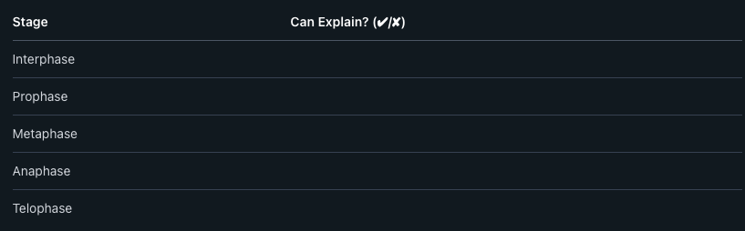
Short activities like matching terms with their definitions also help reinforce learning.
Summative Assessment Approaches
Summative assessments check a student’s understanding after the unit is complete. Common examples include written tests with diagrams, multiple-choice, and short-answer questions.
Teachers may ask students to label the phases of cell division on a diagram. Another approach is having students compare mitosis and meiosis using a table:
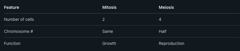
Projects such as creating models or presentations on cell division give students the chance to show knowledge in different ways.
Grading rubrics with clear criteria help ensure scoring is fair and consistent.
Encouraging Student Reflection
Student reflection helps learners think about what they know and what they still need to study. Simple writing prompts ask students to explain, in their own words, how and why cells divide.
Journals are a useful tool. Students can record what they learned each day and write questions about unclear concepts, such as the role of spindle fibers.
Teachers might use one-minute papers, where students answer a prompt like, “What was the most confusing part of today’s lesson on cell division?” This guides instruction for the next class.
Self-assessment checklists help students track their own progress, recognize strengths, and identify areas for review.
Common Challenges and Misconceptions
Students often mix up the details of mitosis and meiosis, such as what happens to DNA and chromosome numbers. They may also misunderstand important words used during cell division lessons, which can make learning harder.
Addressing Student Preconceptions
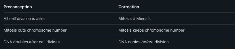
Many students think that all cells divide the same way. They may not realize that mitosis and meiosis have different purposes and outcomes.
It is common for students to believe that mitosis halves the chromosome number, but mitosis keeps the chromosome number the same. In meiosis, however, the chromosome number is halved to produce sex cells.
Another confusion is that DNA is always doubled after division, but DNA is actually copied before cells divide, so each new cell gets the correct amount. Teachers can help by using diagrams showing each step where DNA changes.
Misconceptions Table:
Clarifying Key Terms
Chromosome, chromatid, and chromatin are often confused. Students may mix up these terms, not realizing that:
Chromosomes are X-shaped during cell division.
Chromatids are each half of the X-shaped chromosome.
Chromatin is DNA in its loose, stringy form before cell division.
The terms haploid and diploid can also cause problems. Haploid cells have half the usual chromosome number, while diploid cells have the full set. It helps to use side-by-side examples with pictures for each word.
Clear definitions and simple visuals make it easier for students to match terms to each step in mitosis and meiosis. Using flashcards or short quizzes on these words can reinforce meaning and help students avoid future confusion.
Frequently Asked Questions
Teaching cell division works best when lessons use clear language, hands-on activities, and simple visuals. Interactive tools, active participation, and relatable examples help students understand mitosis and meiosis.
What are effective strategies for teaching the cell division process?
Teachers often use diagrams, videos, and drawing activities to show each step of cell division. Comparing the process to real-life events, such as baking or organizing books, can help make the topic familiar. Breaking stages into small steps lets students learn each part before moving on.
How can interactive worksheets enhance the learning of cell division?
Interactive worksheets give students practice labeling diagrams, answering short questions, and matching terms. They help reinforce vocabulary like prophase and metaphase. Digital worksheets sometimes include quizzes and interactive images for more active learning.
Can you suggest engaging project ideas that illustrate mitosis and meiosis?
Students can create cell division models using clay or craft materials. They can also act out each phase with classmates or use comic strips to draw the stages. Making stop-motion videos with simple materials lets students explain each phase step-by-step.
What is a comprehensive lesson plan for teaching cell division in Grade 8?
A Grade 8 lesson plan may start with a simple introduction to cell division. Teachers often show a video, then guide students through drawing or building each phase of mitosis and meiosis. Worksheets and group discussion at the end help with review and questions.
How important is incorporating a mnemonic for facilitating the understanding of cell division?
Mnemonics help students remember the order of stages, like "PMAT" for prophase, metaphase, anaphase, and telophase. Using familiar phrases makes it easier to recall the steps during lessons or tests. Teachers may ask students to invent their own mnemonics based on class topics.
What are some captivating activities to motivate students during lessons on the cell cycle?
Class games like trivia or card matching can make the cell cycle more engaging. Group work, where students build models of dividing cells, encourages teamwork. Watching animated clips or doing quick classroom experiments with microscopes can also spark interest.


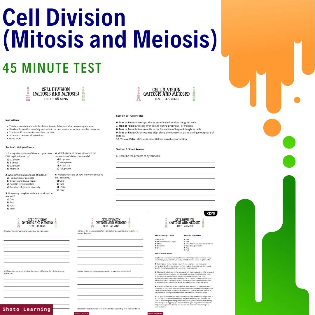


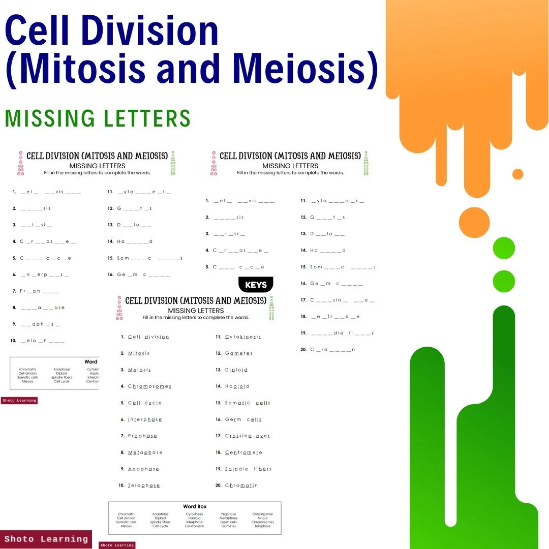


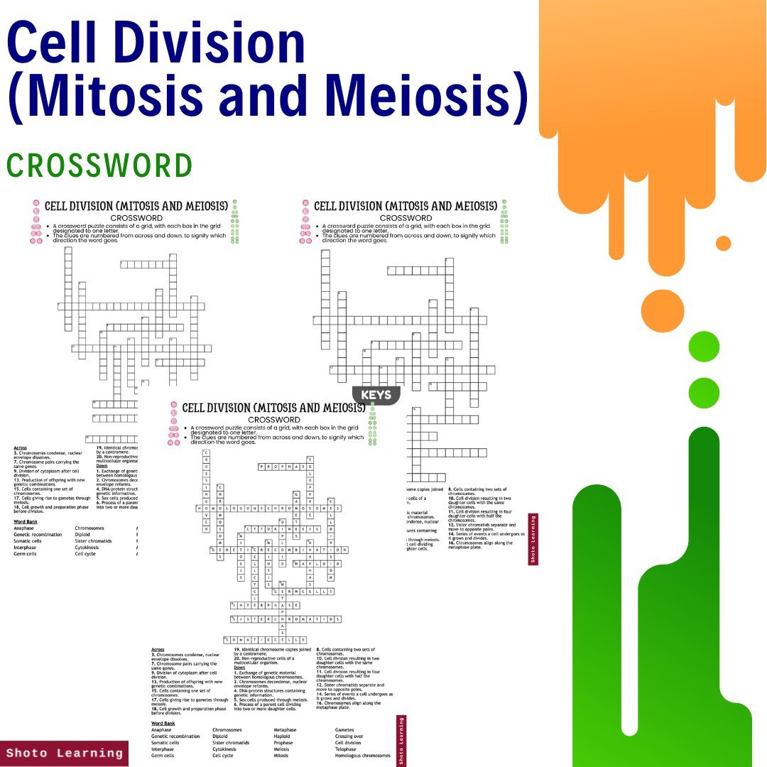


Unlock the mysteries of cellular reproduction and master the concepts of mitosis and meiosis with our Cell Division Mastery Bundle. This comprehensive collection of educational resources provides a wealth of engaging materials to enhance learning and understanding of cell division.
Key Features:
Assignments: Embark on 10 stimulating assignments covering diverse topics, encouraging critical thinking and practical application of knowledge.
Bingo - 30 Bingo Cards: Make learning fun with our interactive bingo game, featuring 30 cards filled with key terms and concepts related to cell division.
Crossword: Challenge your knowledge with a crossword puzzle that tests your understanding of mitosis and meiosis terminology.
Cryptogram: Decode secret messages related to cell division with our cryptogram activity, adding an element of mystery to your learning experience.
Debate: Engage in lively discussions and debates on cell division topics, fostering critical thinking and analytical skills.
Experiments: Explore hands-on experiments illustrating fundamental principles across various disciplines, fostering deeper understanding and engagement.
Fill in the Blank: Reinforce your understanding of cell division concepts with fill-in-the-blank exercises that provide practice and review.
Flashcards: Study on the go with our flashcards, featuring essential terms and definitions related to mitosis and meiosis.
Free ABC Order: Practice organizing key terms alphabetically with our free ABC Order worksheet, reinforcing vocabulary skills.
Free Story Book: Dive into the world of cellular reproduction with our free storybook, offering an engaging narrative on the processes of mitosis and meiosis.
Free Poster: Decorate your learning space with our free poster, providing a visual overview of cell division processes and stages.
Lesson Plan: Dive deep with our detailed lesson plan, complete with engaging activities and discussion topics.
Matching: Test your knowledge of cell division terminology with our matching activity, pairing terms with their corresponding definitions.
Missing Letters: Challenge yourself with our missing letters worksheet, filling in the blanks to complete cell division-related words.
Multiple Choice: Assess your understanding of cell division concepts with multiple-choice questions that cover a range of topics.
PowerPoint: Explore the intricacies of mitosis and meiosis with our informative PowerPoint presentation, featuring detailed explanations and visual aids.
Questions and Answers: Test your knowledge with a set of questions and answers that cover key concepts in cell division.
Reading Comprehension: Sharpen reading skills with engaging passages, perfect for students and educators alike.
Scramble: Unscramble cell division-related words with our scramble worksheet, sharpening your vocabulary and spelling skills.
Test (45 Minutes): Evaluate your understanding of mitosis and meiosis with a 45-minute test that covers essential topics in cellular reproduction.
True or False: Determine the accuracy of statements related to cell division with our true or false activity.
Word Search: Have fun while reinforcing vocabulary with our word search puzzle, featuring cell division-related terms hidden within.
Writing Prompts: Stimulate critical thinking and creativity with a set of writing prompts that explore various aspects of cell division.
Embark on a journey of discovery and mastery with our Cell Division Mastery Bundle. Whether you're a student, educator, or lifelong learner, these engaging resources provide valuable tools and insights to deepen your understanding of mitosis and meiosis.
Pages: 135 pages
Order your copy today!