
Teaching Cell Structure and Function: Strategies for Simplifying Complex Biology Concepts
Understanding cell structure and function can be tough for many students, but it is a key topic in science classes like AP Biology. Teachers can make this subject easier by using clear visuals, real-world examples, and hands-on activities. These strategies help students break down complex information and see how it applies to life around them.
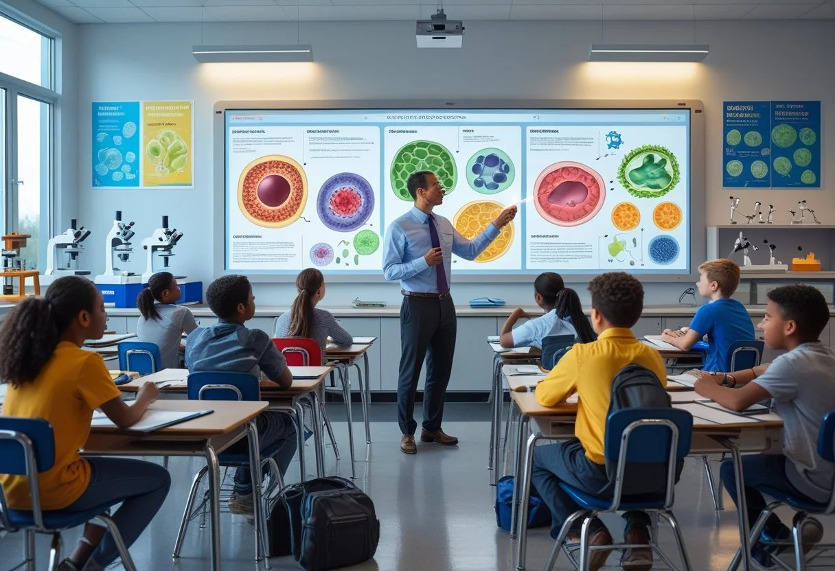
Cells are the basic building blocks of all living things. Learning about their parts and how they work sets the stage for understanding bigger ideas in biology, from growth to how the body stays healthy. Making cell lessons active and engaging supports stronger learning and critical thinking.
Key Takeaways
Cells are the foundation for all living organisms.
Teaching should use active and clear methods for better understanding.
Real-life examples connect cell biology to students’ everyday experiences.
The Cell as the Fundamental Unit of Life
Cells are the smallest unit that show all the characteristics of life. Everything that is alive, from the simplest bacteria to the largest animals, is made of cells.
Defining the Cell Theory
The cell theory is a key idea in biology. It is based on three main points:
All living things are made of cells.
The cell is the basic structural and functional unit of all life.
All cells come from existing cells.
This theory began in the 1800s. Scientists like Matthias Schleiden, Theodor Schwann, and Rudolf Virchow helped develop it. Today, every biologist relies on cell theory to understand how living organisms work.
Cell theory explains not just what cells are, but how life continues and grows. It shows why cells are so central to biology.
Cells as Building Blocks of Life
Cells are often called the building blocks of life. Each cell has structures that help it live, grow, and do its job.
Cell membrane: Thin layer that covers the cell and keeps everything together.
Cytoplasm: Jelly-like fluid inside the cell.
Nucleus: The part that holds genetic information (found in plant and animal cells).
Organelles: Little parts in the cell with specific jobs. Examples: mitochondria and ribosomes.
Cells make up all the parts of living organisms. Even our skin, muscles, and bones are just groups of cells working together.
Cells can look and act very different from each other. For example, blood cells carry oxygen, while nerve cells send messages.
Differentiating Between Unicellular and Multicellular Organisms
Living things can be unicellular (one cell) or multicellular (many cells).
Unicellular organisms—like bacteria and some algae—are made up of a single cell. This cell does all the work: eating, growing, responding to changes, and reproducing.
Multicellular organisms have many cells of different shapes and jobs. In these organisms, each cell is often specialized. For example, humans, dogs, and trees are all multicellular. Their cells work together in groups called tissues and organs to do specific tasks.
Both unicellular and multicellular life forms show how important the cell is as the structural unit of living organisms. The main difference is in how complex the organism becomes as the number of cells increases.
Types of Cells: Prokaryotic and Eukaryotic
All living things are made up of cells, but not all cells are the same. Cells can be grouped into two main types based on their structures and functions.
Characteristics of Prokaryotic Cells
Prokaryotic cells are the simplest and oldest type of cells. Bacteria are the most well-known prokaryotes, but archaea are another group. These cells do not have a nucleus. Instead, their genetic material floats freely in the cell.
Prokaryotic cells are usually much smaller than eukaryotic cells. They lack membrane-bound organelles, which means they don’t have structures like mitochondria or endoplasmic reticulum. Most prokaryotes have a cell wall that helps them keep their shape and protects them.
A typical prokaryotic cell has:
A single circular DNA molecule (not inside a nucleus)
Cell membrane and cell wall
Cytoplasm
Sometimes, small rings of DNA called plasmids
Prokaryotic cells carry out many important jobs in nature. Bacteria, for example, help break down waste and cycle nutrients in ecosystems.
Characteristics of Eukaryotic Cells
Eukaryotic cells are larger and more complex than prokaryotic cells. They have a true nucleus, where genetic material is stored and protected by a membrane. This makes eukaryotic cells unique.
These cells also contain many membrane-bound organelles. Each organelle has a specific job. For example, mitochondria make energy, and the endoplasmic reticulum helps make proteins and fats.
Eukaryotic cells make up:
Animals (including humans)
Plants
Fungi
Protista
Plant cells have cell walls and chloroplasts for photosynthesis. Animal cells do not have cell walls or chloroplasts but have centrioles. Fungi and protists are eukaryotes too, each with their own unique features.
Comparative Analysis of Cell Types
The table below highlights the main differences between prokaryotic and eukaryotic cells:
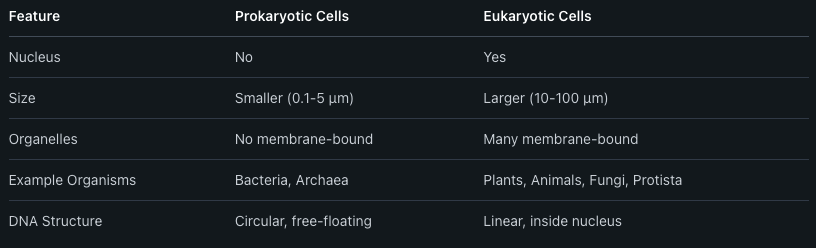
Prokaryotic cells are found mostly in single-celled microorganisms like bacteria. Eukaryotic cells make up more complex organisms, including plants, animals, fungi, and protists. This division helps scientists understand how life is organized and how cells carry out their functions.
Key Cell Structures and Their Functions
Cells are organized into different parts that each play vital roles. Some parts act as barriers, others give support, and some control the flow of information.
Plasma Membrane and Cell Membrane
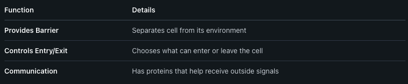
The plasma membrane, often called the cell membrane, surrounds every cell and keeps it together. This membrane is mainly made of a double layer of fats called phospholipids and has proteins stuck in it.
The plasma membrane controls what enters and leaves the cell. It lets in water, oxygen, and nutrients, but keeps out unwanted substances. Signals from the outside world are received through special proteins on the membrane.
A simple table for what it does:
Without a healthy membrane, the cell can’t survive.
Cytoplasm and Organelles Overview
The cytoplasm fills the space between the cell membrane and the nucleus. It is a jelly-like fluid where many cell activities happen. Floating in the cytoplasm are organelles, which act like small organs with different jobs.
Key organelles include:
Mitochondria: Make energy for the cell.
Ribosomes: Build proteins from amino acids.
Endoplasmic Reticulum (ER): Moves materials around. It can be rough (with ribosomes) or smooth (without ribosomes).
Golgi Apparatus: Packages and sends proteins to the right place.
Lysosomes: Break down waste.
These parts work together to keep the cell alive and functioning.
Nucleus and Genetic Material
The nucleus is the main control center of the cell. It is surrounded by its own double membrane which protects it. Inside the nucleus, genetic material is stored.
This genetic material is DNA (deoxyribonucleic acid). DNA holds instructions for making all proteins the cell needs. When needed, parts of DNA are copied into RNA (ribonucleic acid), which then helps build proteins.
DNA is organized into structures called chromosomes. Human cells have 46 chromosomes in their nucleus. Without the nucleus and its genetic material, the cell would not know how to grow, survive, or reproduce.
Specialized Organelles in Plant and Animal Cells
Many cell functions depend on special organelles. Differences in these structures help plant and animal cells carry out their unique roles in living things.
Chloroplasts and Photosynthesis
Chloroplasts are found in plant cells and some algae, but not in animal cells. They contain green pigments called chlorophyll, which allow plants to capture light energy from the sun. This energy is used during a process called photosynthesis.
Photosynthesis is the way plants make their own food. Chloroplasts help turn carbon dioxide and water into glucose, a type of sugar, while releasing oxygen as a byproduct. This happens when light energy is captured by chlorophyll.
Key Points:
Only plant cells have chloroplasts.
Chlorophyll absorbs light energy.
Plants produce glucose and oxygen from carbon dioxide and water.
Table: Summary of Chloroplast Function

Mitochondria: The Cell’s Powerhouse
Mitochondria are organelles in both plant and animal cells. Their main job is to make energy that the cell can use. They do this by breaking down glucose and oxygen to create ATP, the main energy source for cells.
This process is called cellular respiration. The mitochondria are sometimes called the cell’s "powerhouse" because they supply the cell with almost all the energy it needs.
Important Facts:
Both plant and animal cells have mitochondria.
They use glucose and oxygen to make ATP.
ATP powers most activities in the cell.
Vacuoles, Lysosomes, and Golgi Apparatus
Plant and animal cells both have vacuoles, but they are much larger in plant cells. Plant cell vacuoles mainly store water and help support the cell’s shape. Animal cell vacuoles are smaller and used for storing different things.
Lysosomes are found mostly in animal cells. They contain chemicals that break down waste, old cell parts, or food.
The Golgi apparatus is found in both plant and animal cells. It works like a post office, packaging and shipping proteins and other important molecules where they are needed.
Quick Comparison:
Vacuoles: Larger in plant cells, store water.
Lysosomes: Break down waste (mainly in animal cells).
Golgi apparatus: Processes and ships cell materials.
Cell Processes and Function
Cells perform many chemical processes to stay alive and function properly. They make proteins, move substances across their membranes, and send signals to other cells. These activities help cells get energy, use nutrients, and respond to their environment.
Protein Synthesis: Transcription and Translation
Protein synthesis is how cells make proteins. This process has two main steps: transcription and translation. During transcription, information from DNA is copied into a molecule called mRNA. This happens in the nucleus.
Next is translation, which takes place in the cytoplasm. The mRNA moves out of the nucleus and attaches to a ribosome. The ribosome reads the mRNA and puts together amino acids in the correct order to form a protein.
The table below shows the steps:

Proteins built through these steps help with growth, repair, and everyday cell activities.
Cell Membrane Transport and Semi-Permeability
The cell membrane is a semi-permeable barrier that surrounds every cell. This means it lets some things pass through but blocks others. Cells use different ways to move substances in and out.
Types of membrane transport:
Diffusion: Small molecules move from areas of high to low concentration.
Osmosis: Water moves through the membrane by diffusion.
Active Transport: Cells use energy to move materials against a concentration gradient.
Nutrients such as glucose and ions enter the cell, while waste products leave. Proper transport is crucial for cell health and function.
Cellular Communication and Signaling
Cells must communicate with each other to work as a group. This happens through chemical signals sent between cells. These signals can be hormones or other molecules that attach to receptors on the cell surface.
When a signal binds to a receptor, it triggers changes inside the cell. This can turn genes on or off, change the cell’s behavior, or cause it to take in or release nutrients.
Three main steps in cell signaling:
Signal reception (signal binds to receptor)
Signal transduction (message passed inside the cell)
Cellular response (cell acts based on the message)
Communication helps the body coordinate growth, healing, and responses to changes in the environment.
Reproduction and Growth in Cells
Cells use different ways to reproduce and grow. The process depends on whether the cell is prokaryotic or eukaryotic, and if it is going to make more of itself or create cells for reproduction.
Mitosis and Binary Fission
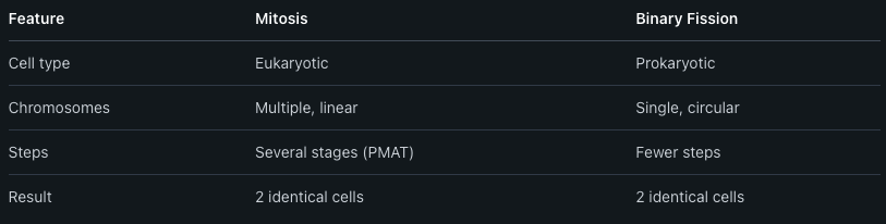
Mitosis is the way most eukaryotic cells divide to make new, identical cells. It lets multicellular organisms grow, repair wounds, and replace old cells. During mitosis, the cell copies its chromosomes so each new cell gets a full set.
Binary fission is how bacteria and other prokaryotes reproduce. It is a simpler process than mitosis. The cell copies its single circular chromosome and then splits into two new cells. Both new cells are almost exactly the same as the original.
Key differences between mitosis and binary fission:
Meiosis in Eukaryotes
Meiosis is how animals, plants, and other eukaryotes make sex cells (like sperm and egg). It has two main cell divisions. The cell starts with chromosomes in pairs, but when meiosis is done, each new cell gets only one chromosome from each pair.
This means each sex cell has half the usual number of chromosomes. When two sex cells come together during fertilization, the right number of chromosomes is restored. This process increases genetic variety because chromosomes get mixed up and exchanged during meiosis.
Meiosis is more complicated than mitosis. It works in two parts called meiosis I and meiosis II. Crossing-over and independent assortment during meiosis help create unique offspring.
Cell Cycle and Chromosome Dynamics
The cell cycle describes the steps a eukaryotic cell goes through as it grows and divides. The main stages are:
Interphase: The cell grows, does its job, and copies its chromosomes.
Mitosis/Meiosis: The cell splits its chromosomes and divides.
Cytokinesis: The cell finishes dividing into two distinct cells.
During these stages, chromosomes are very important. In interphase, DNA is loose. Before cell division, chromosomes become visible and pair up. The cell checks for mistakes before dividing, which helps prevent problems.
Cells must carefully separate chromosomes so each new cell gets the correct amount. If chromosomes are not divided correctly, it can cause errors that may lead to diseases, like cancer. Understanding these steps helps explain how living things grow, repair, and reproduce.
Tools and Techniques for Teaching Cell Structure
Teaching cell structure is more effective with the right tools and teaching methods. Using visuals, real data, and structured discussion helps students understand small cell parts and their roles.
Microscope Use and Magnification
Microscopes are basic tools in biology labs. They let students see parts of cells like the nucleus and membranes. Using magnification, students can compare the detail seen with a light microscope to that of an electron microscope.
An electron microscope offers higher magnification and clearer images compared to light versions. This makes it possible to see smaller structures like ribosomes.
A table of comparison:

Hands-on microscope use gives students practical experience and helps make abstract ideas more real.
Interpreting Experimental Data
Learning how to read and interpret experimental data is key in understanding cell structure. Students look at images, graphs, or charts showing things like the effect of salt or sugar on cells.
Teachers can provide data sets from real experiments. Students learn to spot patterns and make sense of results. For example, they may look at how plant cells change shape in different solutions.
Lists help students organize their thoughts:
Review experiment goals
Look for patterns
Form simple explanations
Encouraging questions and discussions about what the data shows makes the learning process active, not passive.
Mind Mapping Strategies
Mind mapping helps students organize and link ideas. Tools like EdrawMind let them build a cell concept map where terms like “nucleus,” “membrane,” and “mitochondria” branch off from a central cell.
A digital mind mapping tool is useful for building step-by-step diagrams. Students can create a biology concept map to break down the cell’s parts and functions.
Benefits of mind mapping:
Makes complex information clear
Shows connections between cell parts
Aids memory and recall
Using visuals in mind maps helps students understand both the “big picture” and the small details.
Collaborative Learning Platforms
Collaborative learning platforms allow students to work together online and in real-time. They can discuss cell structure, share images or notes, and build group projects from any location.
Features often include chat, file sharing, and real-time collaboration on documents. Students can use these platforms to compare findings from microscope labs or group mind mapping activities.
Collaborative tools make it easy to split tasks. For example, one student uploads microscope images while another adds notes. This division of work supports teamwork and makes cell structure more understandable.
Connecting Concepts to Everyday Life
Learning about cell structure and function helps students see the direct connection between biology and the world around them. Knowledge of human cells, how the body fights disease, and where cells appear daily makes abstract science easier to understand and remember.
Cells in Human Health and Disease
Human health relies on trillions of cells working together. For example, skin is made up of many layers of cells that protect the body from germs and injury. If skin cells get damaged, wounds may not heal well or infections may develop.
White blood cells play a big role in defending the body. They find and destroy germs like bacteria and viruses. When someone feels sick, it is often because white blood cells are fighting hard to keep the body safe.
Many diseases are caused when certain cells stop working right. For example, cancer happens when cells begin to grow in an uncontrolled way. Diabetes is related to problems with insulin-producing cells in the pancreas. Knowing about cell structure helps explain how these diseases develop and why treatments target specific cells.
Examples of Cells in Daily Life
Cells are not just in science labs—they are all around, even if they can't be seen. When a person skins their knee, skin cells work to close the cut. Holding something cold or hot, nerve cells send signals to the brain to sense temperature and pain.
A table of everyday examples can help:
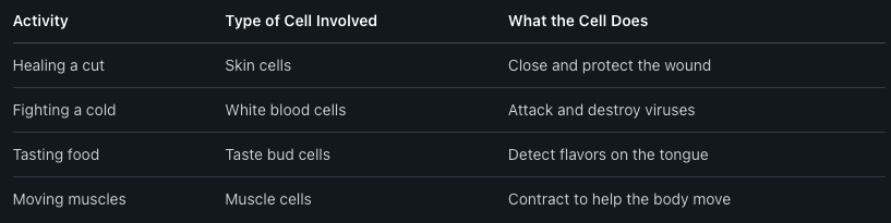
Seeing how cells work in daily life makes biology more real. It encourages people to connect what they learn in class to their own experiences, from feeling sick to playing sports.
Frequently Asked Questions
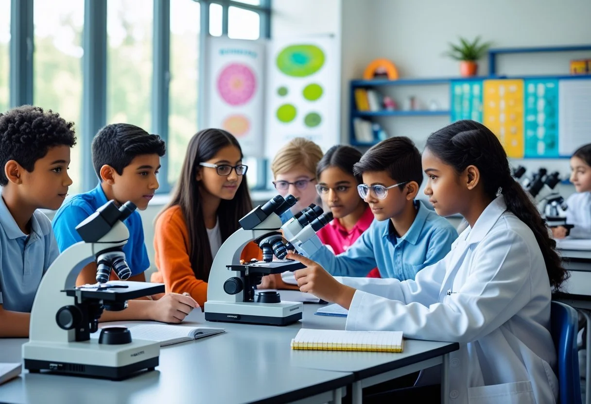
Teachers often use models, visual aids, and interactive technology to help students understand complex ideas about cells. Engaging activities, hands-on experiments, and creative lessons can break down big science concepts in ways students can remember.
What are effective hands-on activities for teaching cell structure and function?
Students can build 3D models of plant and animal cells using clay, gelatin, or candies. Microscopy labs where they look at real cells help them see the differences between prokaryotes and eukaryotes. Coloring diagrams and making cell organelle flipbooks also support learning.
What interactive simulations can help students understand cell structures?
Online tools like BioMan Biology and ExploreLearning Gizmos let students explore interactive cell models. These programs often allow students to click on organelles, change cell types, and run simple experiments. Many simulations show how organelles work together in the cell.
How can cell organelle functions be taught engagingly to high school students?
Teachers may use games like cell structure bingo or matching activities to connect names with organelle jobs. Role-playing, where students act out organelle functions, can help them remember each part’s job. Short videos and interactive apps keep lessons fresh and interesting.
How can worksheets be used to reinforce the learning of cell parts and functions?
Labeling activities on worksheets help students recall organelle names. Fill-in-the-blank exercises and crosswords can reinforce vocabulary about cell functions. Some worksheets ask students to draw their own cells or match organelles to their functions, building stronger connections.
Why is learning about cell functions critical for students?
Understanding cell functions helps explain how living things grow, heal, and react to their environment. It connects to health topics, genetics, and how the human body works. Cell biology provides a foundation for learning about life in more detail later.
What are some creative methods to make cell biology interesting for learners?
Teachers can use songs, cartoon drawing, or storytelling to explain how cells work. Creating posters or digital presentations lets students show cell processes in new ways. Simple experiments with yeast or plant cells give learners a real-world view of cell activity.


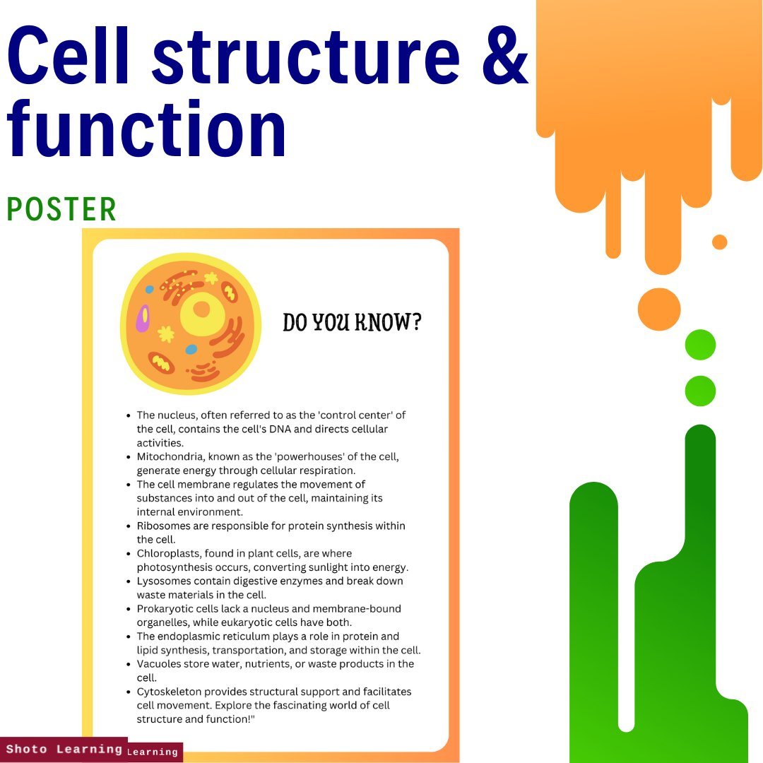
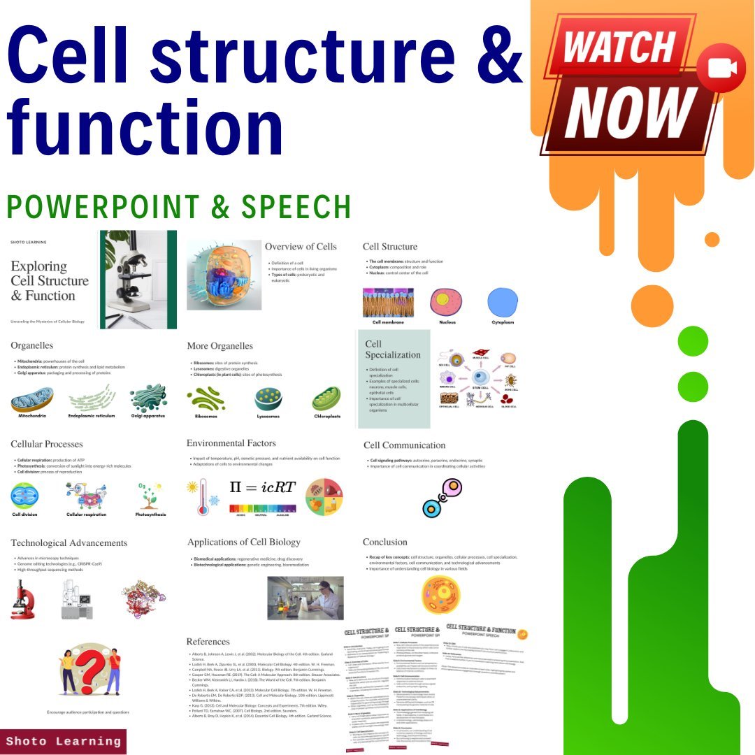
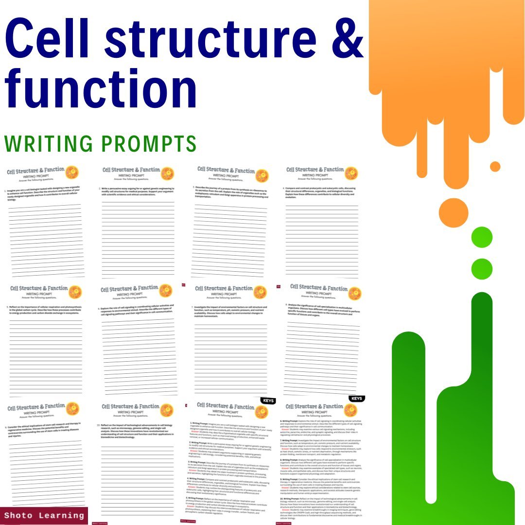

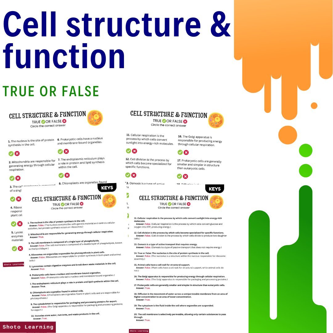

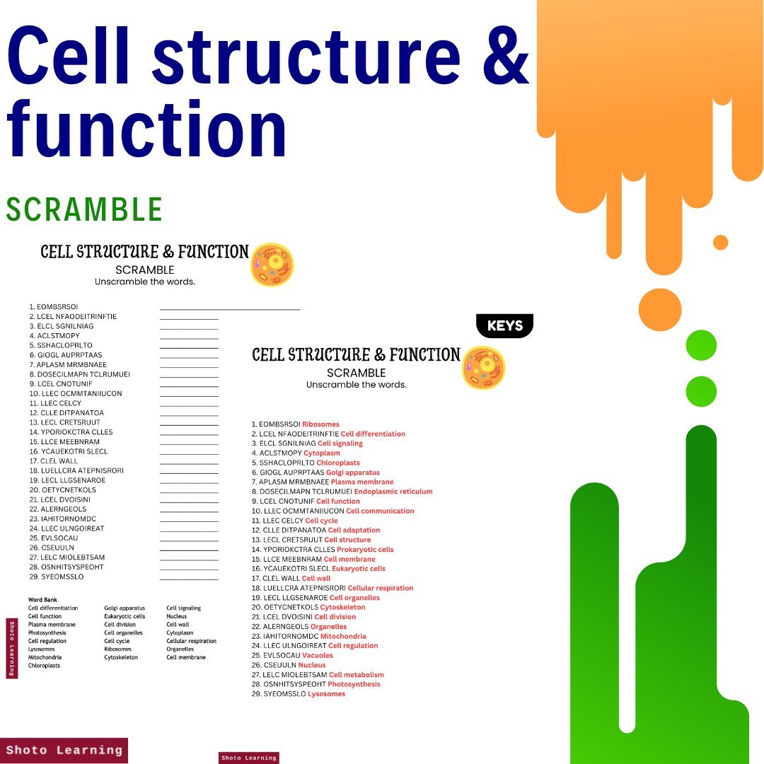

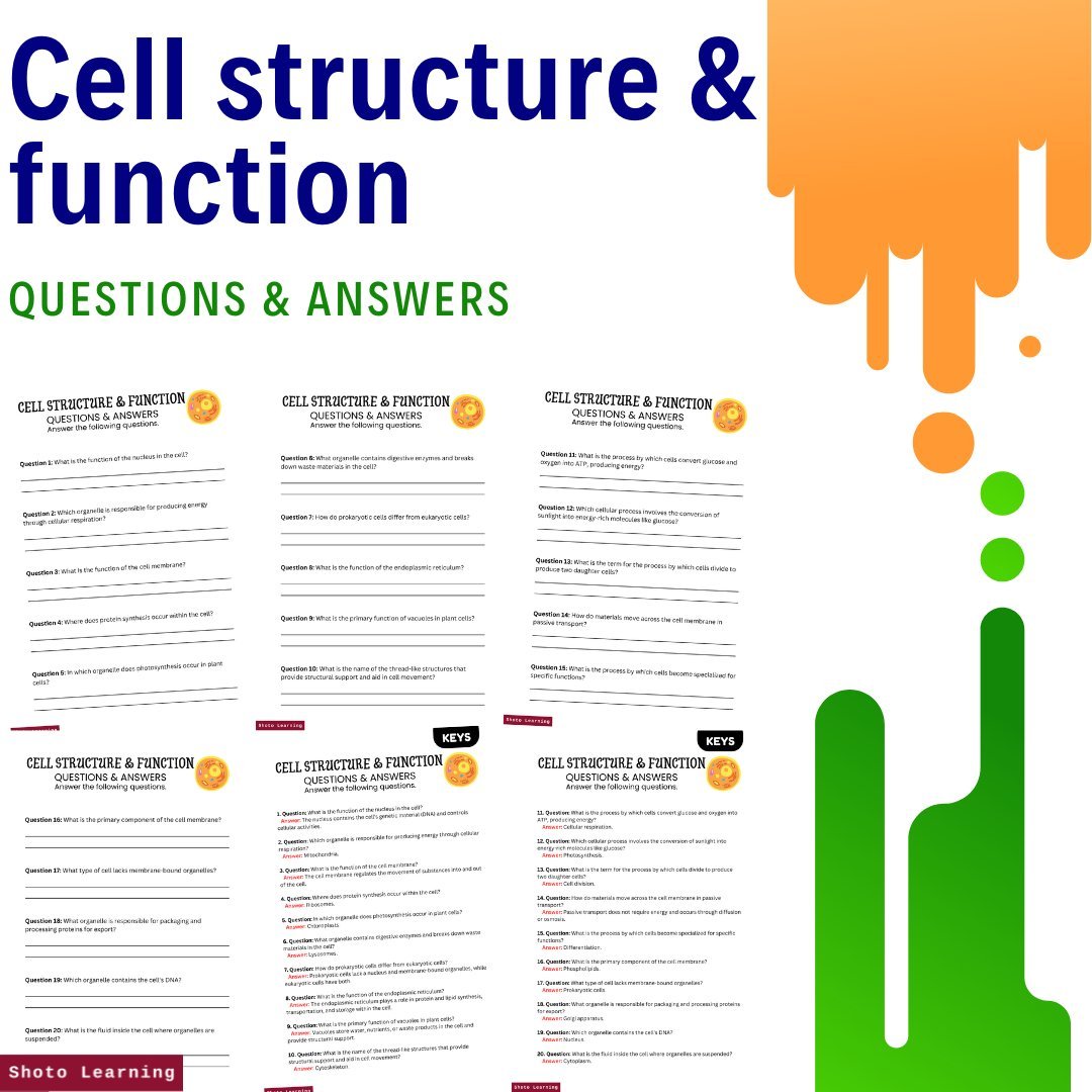
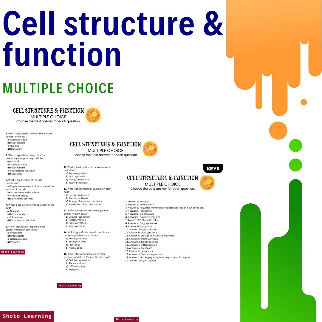
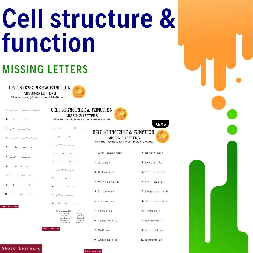
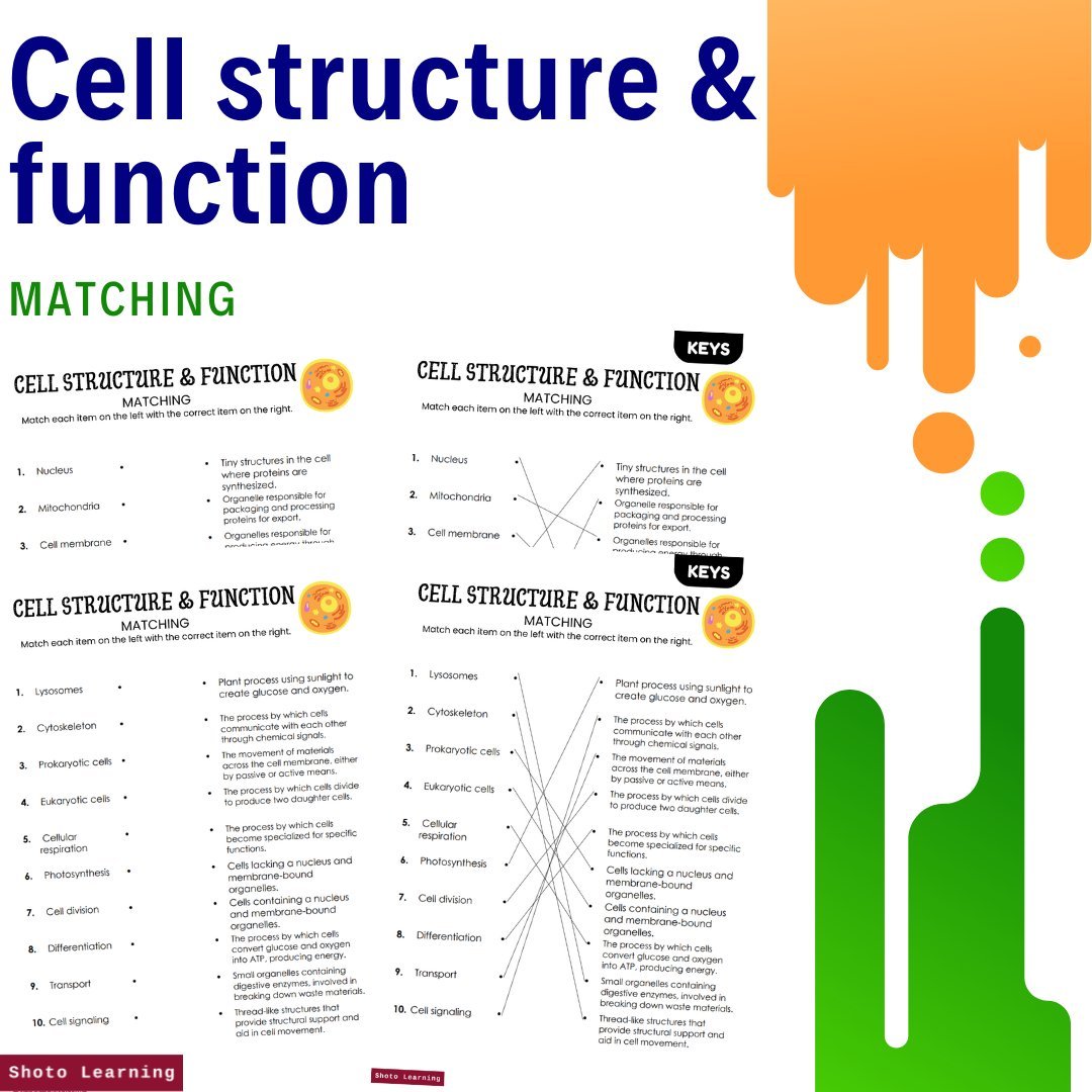

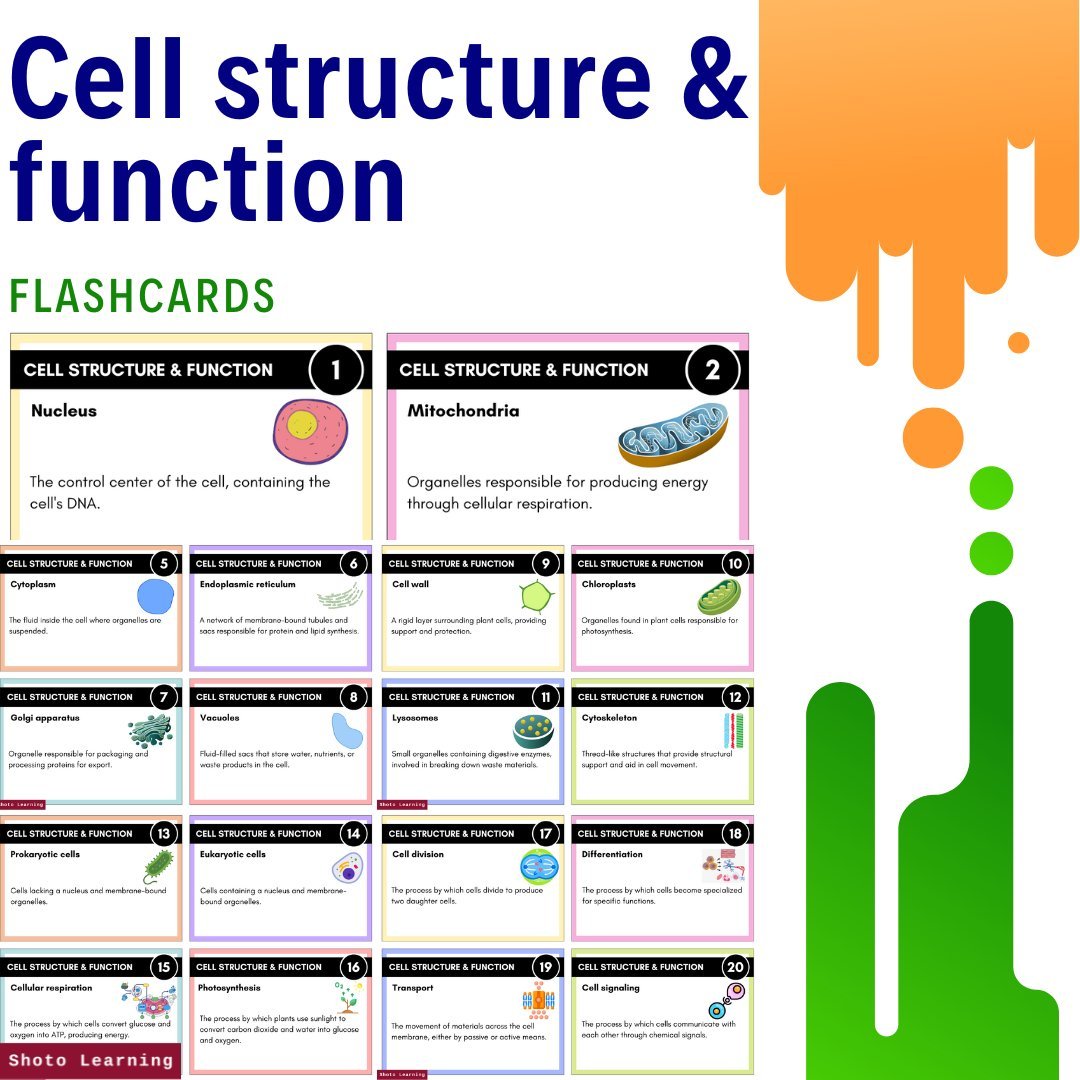


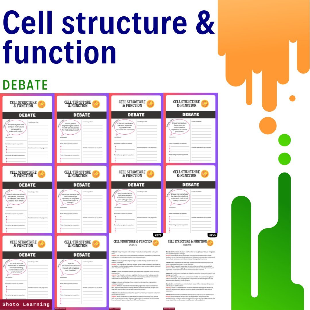


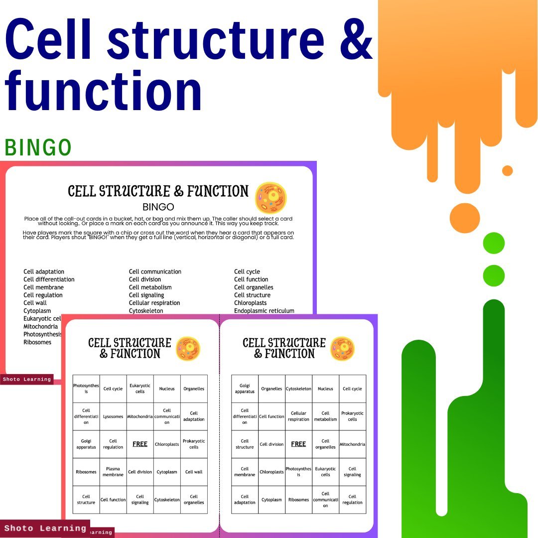

Embark on an immersive journey into the intricate world of cellular biology with our "Cell Structure & Function Mega Bundle," meticulously crafted to ignite curiosity and foster deep understanding. This comprehensive package offers an array of captivating resources, meticulously designed to engage learners and fortify their grasp of fundamental concepts in cellular biology.
Key Components:
1. Dynamic Lesson Plan: Dive into stimulating discussions and activities orchestrated to unravel the mysteries of cell structure and function. Our meticulously crafted lesson plan provides a roadmap for educators, complete with clear objectives, innovative instructional strategies, and insightful assessment recommendations.
2. Varied Assignments and Experiments: Delve into a diverse collection of assignments designed to invigorate learning. From stimulating debates and writing prompts to hands-on experiments and challenging puzzles, our bundle offers an extensive array of activities to cater to diverse learning styles and foster critical thinking.
3. Interactive Learning Tools: Engage in interactive learning experiences with our assortment of dynamic resources, including Bingo, Crossword puzzles, and Flashcards. These immersive tools provide a hands-on approach to reinforce key terminology and concepts, ensuring retention and comprehension.
4. Thought-Provoking Exercises: Stimulate cognitive agility and creativity with thought-provoking exercises such as Cryptogram puzzles and thought-provoking debate topics. These exercises encourage students to think critically, analyze information, and articulate their understanding effectively.
5. Comprehensive Assessment Tools: Evaluate comprehension and mastery with our suite of assessment tools, including comprehensive tests, true or false quizzes, and multiple-choice questions. These meticulously crafted evaluations ensure a thorough understanding of cellular biology concepts.
6. Visual Presentations: Enhance learning experiences with visually rich PowerPoint presentations, meticulously curated to deliver comprehensive lectures or review sessions. Informative slides and engaging visuals serve as invaluable aids in reinforcing key concepts and fostering deeper understanding.
7. Bonus Resources: Access additional resources, including ABC Order worksheets, Label the Parts activities, and Posters, to further reinforce learning and provide additional practice opportunities.
Experience Comprehensive Learning: Our "Cell Structure & Function Mega Bundle" offers an unparalleled opportunity to explore every facet of cellular biology. Whether you're an educator seeking to enrich your curriculum or a student eager to delve into the wonders of biology, this bundle is your ultimate companion for success.
Unlock the mysteries of cellular biology and embark on a transformative learning journey with our "Cell Structure & Function Mega Bundle." From engaging assignments to interactive experiments and thought-provoking reading comprehension, this bundle is your gateway to a deeper understanding of the intricate world of cell structure and function. Whether you're a seasoned educator or an eager learner, seize the opportunity to elevate your understanding and unlock new realms of knowledge with our comprehensive bundle.
Pages: 132 pages
Order your copy today!