
How to Explain Cell Structure and Function to Students Effectively in the Classroom
Understanding cell structure and function can seem tough for students, but with the right approach, anyone can learn the basics. Teaching cell structure and function is easier when the focus is on simple explanations and clear connections between form and role. Students are more likely to stay interested when the lesson includes hands-on activities, real examples, and visual tools.
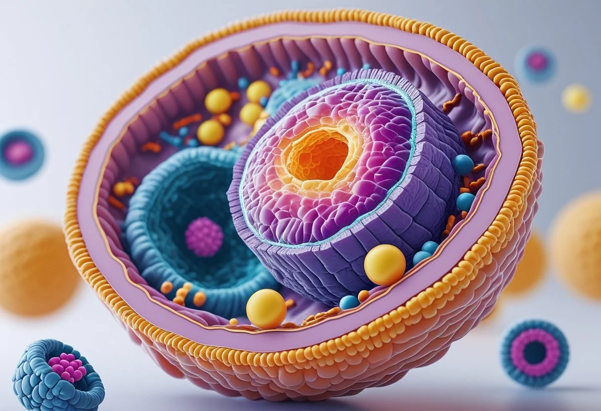
This article offers practical ways to help students grasp key parts of the cell, like the nucleus, cell membrane, and mitochondria. It also explains how different cells—plant, animal, and bacteria—work differently based on their parts. These tips can make teaching cells less overwhelming and more interactive.
Key Takeaways
Focus on clear and simple ways to teach cell structure and function.
Show important differences between types of cells and key organelles.
Use engaging activities and real-life examples to help students understand.
Key Principles of Cell Structure and Function
Cells are the smallest units of life, responsible for all vital activities in organisms. Each part of a cell carries out specific roles that keep the cell functioning.
Understanding the Concept of a Cell
A cell is defined as the basic building block of all living things. Every organism, from tiny bacteria to large plants and animals, is made up of one or more cells.
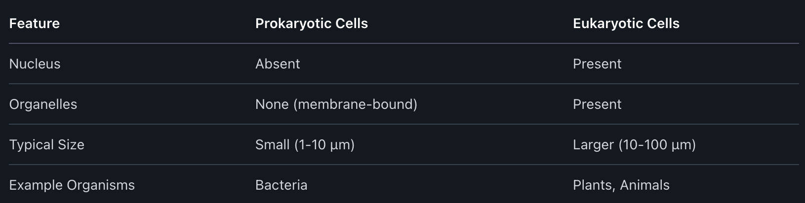
Cells come in two main types: prokaryotic (like bacteria) and eukaryotic (like plants and animals). Prokaryotic cells do not have a nucleus, while eukaryotic cells do.
The cell theory states three main points:
All living things are made of cells.
The cell is the smallest unit that can carry out life’s processes.
All cells come from existing cells.
Cells can exist alone or within groups in multicellular organisms. This ability to form different life forms makes understanding cells important to biology.
Importance of Cell Structures in Life
Each cell has key structures called organelles, which are small parts inside the cell with special jobs. For example, the nucleus stores genetic information, the mitochondria makes energy, and the cell membrane controls what goes in and out.
The shape and parts of a cell have a direct effect on how it works. For instance, muscle cells contain extra mitochondria to meet energy needs, while plant cells have a rigid cell wall to help the plant stand upright.
These main cell organelles include:
Nucleus: Controls cell activities.
Cytoplasm: Gel-like fluid where organelles float.
Mitochondria: Provides energy.
Cell membrane: Protects and regulates entry/exit.
Chloroplasts (in plants): Make food using sunlight.
Understanding what each part does helps students see how cells support life.
Link Between Structure and Function
The way a cell looks and what parts it has help decide what it can do. For example, red blood cells are shaped like discs, making it easier for them to carry oxygen.
Nerve cells have long extensions to send signals quickly. Plant cells have a tough cell wall to help them stay strong and take in water.
This relationship, often called the structure-function principle, explains why cells of different jobs have different shapes or organelles.
Below is a simple table showing examples:
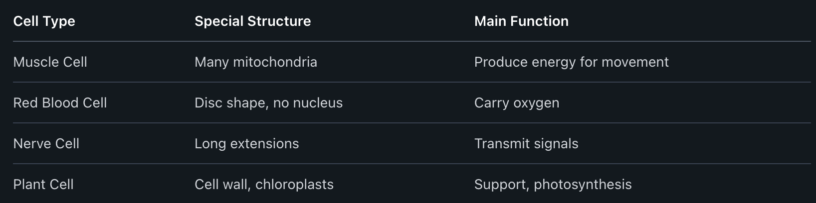
By observing these differences, it is clear that each cell’s design closely matches its role in keeping the organism healthy.
Explaining Cell Theory
Cell theory is a basic idea in biology that helps explain what living things are made of and how they work. It describes facts about cells, important discoveries, and how this knowledge shapes what people know about all living organisms, including bacteria.
Historical Development and Key Scientists
Cell theory began in the 1800s after scientists started using microscopes to look at tiny parts of plants and animals. Robert Hooke was the first to use the word “cell” in 1665. He looked at cork under a microscope and saw small, box-like shapes.
A few years later, Anton van Leeuwenhoek observed single-celled organisms, now known as bacteria and protozoa, in pond water. In the 1830s, Matthias Schleiden and Theodor Schwann stated that plants and animals are made of cells. Rudolf Virchow added to their work in 1855 by saying that new cells come from other living cells.
These scientists’ work built the foundation for cell theory and changed how people think about living things.
Core Statements of Cell Theory
Cell theory has three main ideas. These are known as the core statements:
All living things are made of one or more cells.
The cell is the basic unit of structure and function in living things.
All cells come from pre-existing cells.
These statements apply to all living organisms, from simple bacteria to complex plants and animals. Cells carry out life’s basic activities, like making energy and removing waste. No living thing can exist without cells.
Teachers often show pictures or use models to explain these three points. It helps students see how even the smallest bacteria share the same building blocks as larger organisms.
Modern Cell Theory in the Classroom
Modern cell theory includes both old and new ideas about cells. Today, scientists know that all cells share common features like cell membranes, DNA, and the ability to use energy. Lessons may highlight how both multicellular and single-celled organisms, such as bacteria, perform the functions needed for life.
In the classroom, educators use diagrams, activities, and real examples to make the theory clear. For instance, studying bacteria helps students see how one cell does all the work needed to stay alive. Interactive lessons, group projects, and simple microscopes can help students connect cell theory to real life.
Teachers find that breaking down complex ideas and focusing on the basics helps students understand how important cells are to every living thing. Through these approaches, students learn to apply cell theory to the world around them.
Types of Cells: Prokaryotic and Eukaryotic
Cells come in two main forms, each with unique structures. These types include prokaryotic cells and eukaryotic cells, found in a wide range of living things from bacteria to plants and animals.
Differences Between Prokaryotic and Eukaryotic Cells
The biggest difference is that prokaryotic cells do not have a nucleus, while eukaryotic cells do. In prokaryotic cells, such as bacteria, the DNA floats in the central part of the cell called the nucleoid.
Eukaryotic cells, found in plants and animals, store their DNA inside a nucleus. They also have other membrane-bound organelles like mitochondria and, in plant cells, chloroplasts.
Prokaryotes tend to be smaller and simpler. They usually have a cell wall, cell membrane, cytoplasm, ribosomes, and sometimes flagella for movement. Eukaryotic cells are larger and more complex. They can have specialized structures that carry out different jobs, making them better suited for life in multicellular organisms.
Examples: Bacteria, Plant, and Animal Cells
Bacteria are classic prokaryotes. They have a simple structure with no nucleus and few internal parts. Their cell walls help them survive in many environments.
Plant cells and animal cells are both eukaryotic. They share many of the same inner parts, such as a nucleus, endoplasmic reticulum, and Golgi apparatus.
Plant cells have a cell wall, chloroplasts for photosynthesis, and a large central vacuole for storage. Animal cells do not have cell walls or chloroplasts, but they often have small vacuoles and unique structures like lysosomes.
Below is a simple list:
Bacteria: Prokaryotic, no nucleus, cell wall always present.
Plant cells: Eukaryotic, nucleus present, have chloroplasts, cell wall present.
Animal cells: Eukaryotic, nucleus present, no cell wall, no chloroplasts.
Fundamental Cell Structures and Their Functions
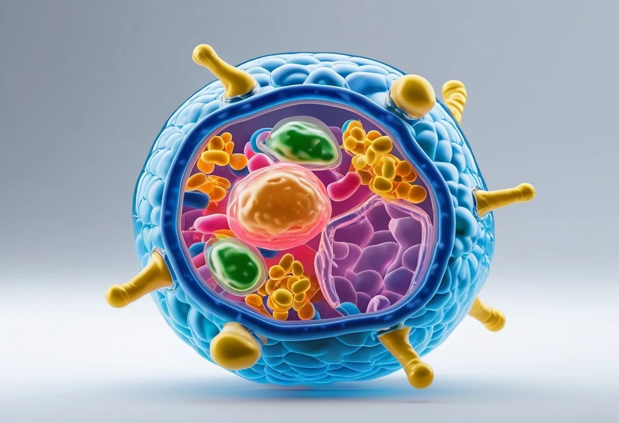
A cell’s basic structures allow it to survive, function, and reproduce. Each major part plays a unique role, from keeping substances in or out to controlling the cell’s activities and protecting it from harm.
Cell Membrane: Role and Selective Permeability
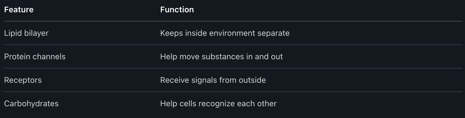
The cell membrane surrounds every cell, forming a flexible barrier between the inside and outside. This barrier is very thin but strong.
It is made mostly of lipids and proteins. The main job of the cell membrane is to control what enters and leaves the cell. This job is called selective permeability.
Some molecules, like water and oxygen, move through easily. Others, like large proteins or ions, need special transport systems. Channels and pumps made from proteins help move these substances.
The cell membrane also helps the cell communicate with other cells. It sends and receives chemical signals, which let the cell respond to changes in its environment.
Cytoplasm: Organization and Activities
The cytoplasm fills the space between the cell membrane and the nucleus. It is a thick, jelly-like fluid.
Most cell activities happen in the cytoplasm. It contains many different organelles, or small structures that do specific jobs. For example, mitochondria make energy, and ribosomes build proteins.
The cytoplasm holds everything in place, so organelles do not float away. It also provides the raw materials and space for chemical reactions that keep the cell alive.
Here are some important organelles found in the cytoplasm:
Mitochondria: Make energy for the cell
Ribosomes: Build proteins
Endoplasmic reticulum: Helps move materials
Nucleus: Control Center and Genetic Material
The nucleus is usually the largest part inside a eukaryotic cell. It acts as the cell’s control center.
The nucleus contains most of the cell’s genetic material in the form of DNA. DNA carries the instructions, called genes, for making all the proteins needed by the cell.
The nucleus is surrounded by a nuclear envelope, which keeps its contents separate from the cytoplasm. Small openings called nuclear pores let messages move in and out.
Key functions of the nucleus include:
Storing genetic information
Sending instructions for making proteins
Directing cell growth and reproduction
Cell Wall: Structure and Protective Functions
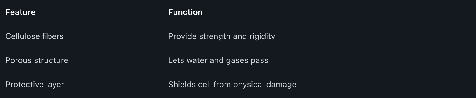
The cell wall is a tough layer found outside the cell membrane in plant, fungus, and some bacterial cells. It is not present in animal cells.
The main job of the cell wall is to give the cell shape and strength. In plants, the wall is made mostly of cellulose, a strong carbohydrate. The cell wall keeps the cell from bursting if too much water enters.
It also protects the cell from harmful chemicals and gives support to help the plant stand upright. Because of the cell wall, plant cells can form rigid structures, like stems and leaves.
Major Cell Organelles and Their Functions
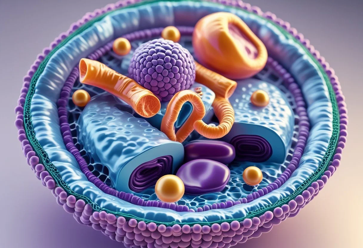
Each cell contains several organelles, which are specialized structures that perform key functions. These organelles help the cell get energy, make proteins, store important materials, and more.
Mitochondria: Cellular Respiration and Energy Production
Mitochondria are known as the "powerhouses" of the cell. They produce energy by breaking down sugars during a process called cellular respiration.
Each mitochondrion has two membranes. The inner membrane is folded to increase the area for chemical reactions. These organelles use oxygen to turn glucose into adenosine triphosphate (ATP), which is the cell's main energy carrier.
Cells that need a lot of energy, like muscle cells, have more mitochondria. In animal and plant cells, mitochondria are essential. Without them, the cell cannot carry out vital activities.
Key facts:

Chloroplasts: Photosynthesis in Plant Cells
Chloroplasts are found only in plant cells and some algae. They are needed for photosynthesis, which allows plants to make their own food from sunlight.
Each chloroplast contains a green pigment called chlorophyll. This pigment traps energy from sunlight. The energy is then used to turn carbon dioxide and water into glucose, a type of sugar. Oxygen is released as a waste product during this reaction.
Chloroplasts have a double membrane like mitochondria. Inside, there are stacks of tiny discs called thylakoids, which increase the surface area for light absorption. Chloroplasts are critical for life on Earth because they produce the oxygen we breathe.
Ribosomes: Protein Synthesis
Ribosomes are the sites where cells build proteins. Proteins are needed for almost every function in the body, such as repairing tissues, making enzymes, and supporting the immune system.
There are two types of ribosomes: free ribosomes float in the cytoplasm, while bound ribosomes attach to the rough endoplasmic reticulum. Both types make proteins by reading instructions from messenger RNA (mRNA).
Unlike organelles with membranes, ribosomes are made mostly of ribosomal RNA (rRNA) and proteins.
Ribosome Highlights:
No membrane
Made of RNA and protein
Make proteins by joining amino acids in the right order
Vacuoles: Storage and Support
Vacuoles are large storage spaces found in the cytoplasm of cells. They have different roles in plant and animal cells.
In plant cells, the central vacuole is very large. It stores water, nutrients, and waste products. By holding water, it also helps the plant cell keep its shape and stay firm. This keeps the plant from wilting.
Animal cells may have smaller vacuoles, which store materials or help in waste removal. Vacuoles are surrounded by membranes that keep their contents separate from the rest of the cell.
Main functions:
Store water and important substances
Support plant cell structure
Remove unwanted materials from the cell
Illustrating Differences Among Plant, Animal, and Bacterial Cells
Plant, animal, and bacterial cells each have important differences in their parts and functions. Understanding how their structures vary makes it easier to see what each cell can do.
Unique Features of Plant Cells
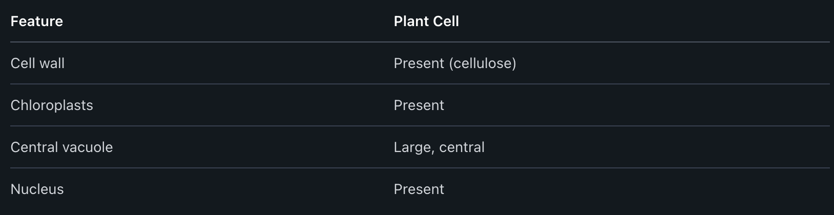
Plant cells are known for having a rigid cell wall made of cellulose. This cell wall helps the cell keep its shape and protects it from damage.
Inside plant cells, there are chloroplasts. These are green organelles that help the plant capture sunlight to make food through photosynthesis. This is a process not found in animal or bacterial cells.
Plant cells also have a large central vacuole. This vacuole holds water, nutrients, and waste. It can take up most of the space inside the cell and helps keep the cell firm.
Other important features include a nucleus that holds genetic material, a cell membrane just inside the cell wall, and many smaller organelles like mitochondria for energy.
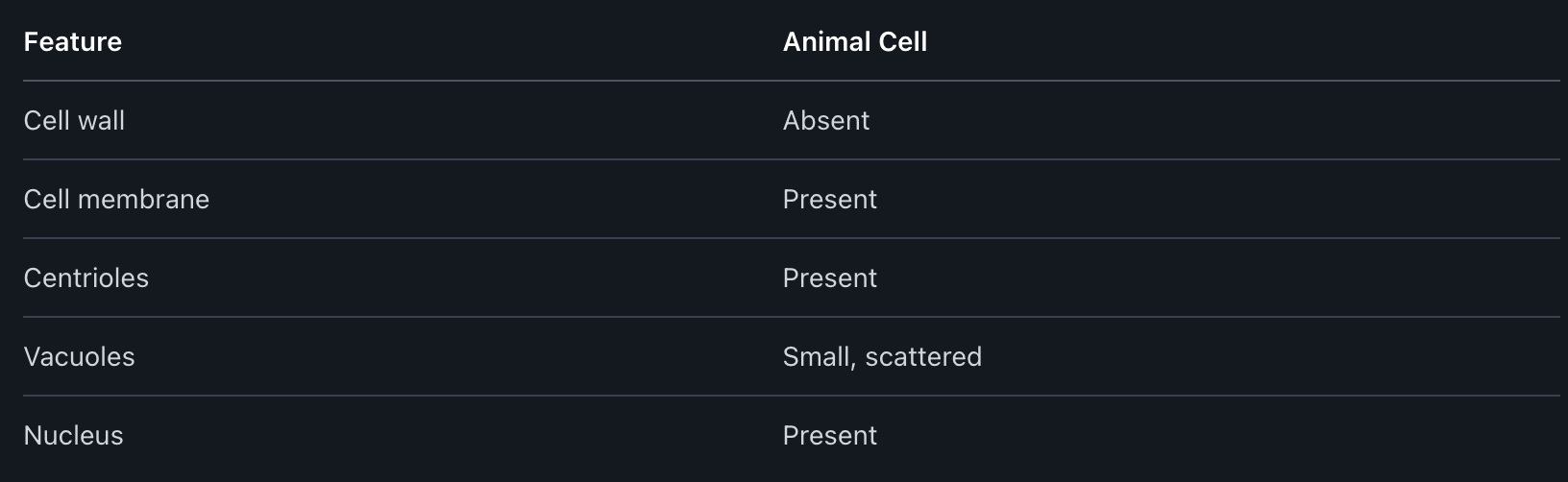
Special Structures in Animal Cells
Animal cells do not have a cell wall. Instead, they are surrounded only by a cell membrane. This membrane controls what enters and leaves the cell and helps the cell stay flexible.
Animal cells have many small vacuoles instead of one large one. They also contain centrioles, which help with cell division.
Unlike plant cells, animal cells do not have chloroplasts or a central vacuole. They also tend to be more rounded or irregular in shape.
Animal cells, like plant cells, have a nucleus, mitochondria, and other organelles. The nucleus directs the cell's actions, while mitochondria provide energy.
Characteristics of Bacterial Cells
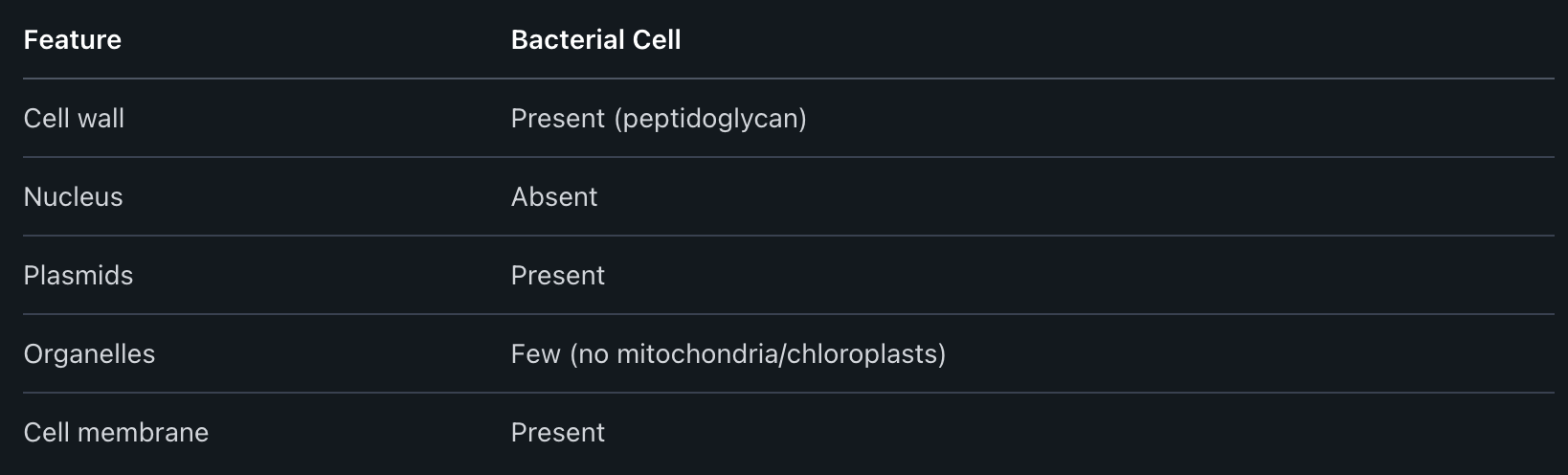
Bacterial cells are prokaryotic, meaning they do not have a true nucleus. Their genetic material is found as a single circular DNA in the cytoplasm.
They have a cell wall, but it is made of a material called peptidoglycan, not cellulose. Some bacteria also have a sticky outer layer called a capsule.
Bacteria lack most of the organelles seen in plant and animal cells. They do not have mitochondria, chloroplasts, or a nucleus. Instead, they have simple structures called ribosomes to make proteins.
The cell membrane is found just inside the cell wall and controls what goes in and out. Some bacteria also have small circles of DNA called plasmids.
Engaging Students in Learning Cell Structure and Function
Bringing cell structure and function to life helps students remember key information. Visual tools, hands-on activities, and real cell observations help students make sense of cell parts and their jobs.
Using Microscopes to Explore Real Cells
Microscopes allow students to see real cells in action. By observing onion skin or cheek cells, they compare plant and animal cells firsthand. Cells Alive and other digital resources also demonstrate what normal, living cells look like, making lessons more relatable.
Teachers can guide students to identify the nucleus, cell wall, or cell membrane while looking under the microscope. A labeled worksheet beside the microscope helps students name each structure as they see it. Group discussions give students the chance to share their observations and correct any mistakes.
Comparing bacteria, plant, and animal cells under the microscope helps students spot differences, like the presence of a cell wall in plant and bacteria cells but not in animal cells. This first-hand look builds understanding of cell structure that a picture can’t provide.
Interactive and Visual Learning Strategies
Interactive lessons boost student engagement. Virtual simulations of cells let students explore organelles by clicking or dragging to learn their names and functions. Cell coloring sheets are effective for helping students visually identify organelles and learn what each part does.
Games, like organelle matching or “cell structure bingo,” encourage group learning and make it easier to remember cell parts. Visual comparisons between plant and animal cells using diagrams or Venn diagrams help students see both differences and similarities at a glance.
Videos showing animations of the inside of a cell can also help students understand how all the cell parts work together. Teachers can pause these videos to discuss the role of the mitochondria, nucleus, or other organelles in real time.
Implementing Hands-On Activities and Models
Model building helps students see the 3D layout of a cell. Using clay, playdough, or even candy, students can form different organelles such as the nucleus, mitochondria, or chloroplast. This approach gives a tactile element to teaching cell structure.
Station activities encourage students to rotate between different “cell tasks,” such as labeling diagrams, matching organelles to their functions, or assembling a model. This keeps lessons active and gives many opportunities for feedback and correction.
A cell scavenger hunt or interactive classroom walk allows students to find organelles or facts posted around the room. This makes learning about cell structure and function more dynamic and less overwhelming, especially for younger students.
Frequently Asked Questions
Cells are the basic building blocks of life. Their internal structures help them work correctly, support life, and react to changes.
What are the primary components of a cell and their respective functions?
Cells have different parts called organelles. Each organelle has a job.
The nucleus controls the cell’s activities. Mitochondria make energy. The cell membrane protects the cell and controls what passes in and out. The endoplasmic reticulum helps build proteins and lipids. Ribosomes make proteins. The Golgi apparatus sorts and ships proteins. Lysosomes break down waste.
How does the structure of cell organelles relate to their roles in the cell?
An organelle’s shape helps it do its job well. For example, mitochondria have folded inner membranes to produce more energy. Ribosomes are tiny, so they can quickly make proteins.
The Golgi apparatus has stacks of membranes that help process and sort proteins. These shapes are directly linked to how each organelle works.
Can you describe the differences between plant and animal cell structures?
Plant cells have a cell wall, which gives them a firm shape. They also have chloroplasts for making food through photosynthesis, and a large central vacuole that stores water.
Animal cells do not have a cell wall or chloroplasts. Their vacuoles are smaller. These differences help each cell do its special functions.
What techniques can be used to teach students about the cell theory effectively?
Teachers can use diagrams, simple models, and hands-on activities. Viewing cells under a microscope lets students see real cell structures. Group discussions and analogies, like comparing a cell to a factory, can help students remember key ideas.
Asking clear questions about the three main points of cell theory helps increase understanding. Breaking information into small parts can keep students from feeling overwhelmed.
In what ways do cell membranes maintain cellular homeostasis?
The cell membrane controls what enters and leaves the cell. It lets in nutrients but keeps out harmful substances. It also removes waste.
This selective control helps the cell keep water, minerals, and other conditions balanced so it can work properly.
How can we use models and diagrams to illustrate cell structure in the classroom?
Using labeled diagrams makes it easier to identify parts of the cell. Physical or digital models allow students to see details in 3D.
Building cell models from clay, paper, or online tools can help students understand how the organelles fit together. These visual aids are useful for both learning and review.


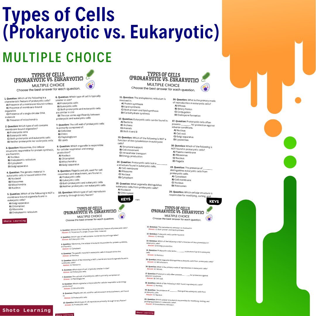


Embark on an exciting journey into the realm of cellular biology with our comprehensive "Cell Biology Activity Bundle," focusing on the comparison between prokaryotic and eukaryotic cells. This bundle features a diverse selection of engaging activities and resources designed to deepen understanding, promote critical thinking, and reinforce key concepts. Whether you're a teacher looking to enhance classroom lessons or a student seeking additional practice, this bundle provides a wealth of educational opportunities. From bingo and crossword puzzles to flashcards and writing prompts, there's something for everyone in this comprehensive bundle.
Bundle Includes:
Assignments: Embark on 10 stimulating assignments covering diverse topics, encouraging critical thinking and practical application of knowledge.
Bingo - 30 Bingo Cards: Enjoy a fun and interactive game of bingo while reinforcing knowledge of prokaryotic and eukaryotic cells. Perfect for classroom activities or study groups.
Crossword Puzzle: Challenge yourself with a crossword puzzle that tests your understanding of cell biology concepts and terminology.
Cryptogram: Decode cryptic messages related to prokaryotic and eukaryotic cells, combining logic and knowledge to unravel hidden meanings.
Debate: Engage in lively debates exploring the differences between prokaryotic and eukaryotic cells, fostering critical thinking and communication skills.
Experiments: Explore hands-on experiments illustrating fundamental principles across various disciplines, fostering deeper understanding and engagement.
Fill in the Blank: Test your knowledge of cell biology with fill-in-the-blank exercises covering various aspects of prokaryotic and eukaryotic cells.
Flash Cards: Reinforce key concepts and terms with flashcards designed for quick review and memorization.
Free ABC Order: Practice alphabetizing cell biology terms with a free ABC order worksheet, promoting vocabulary retention and organization skills.
Free Label the Parts: Identify and label the essential components of prokaryotic and eukaryotic cells with a free labeling worksheet.
Free Poster: Explore intriguing facts about cell biology with a free poster, perfect for classroom display or personal reference.
Lesson Plan: Dive deep with our detailed lesson plan, complete with engaging activities and discussion topics.
Matching: Match descriptions to corresponding cellular structures and functions, reinforcing understanding of key concepts.
Missing Letters: Exercise your problem-solving skills by filling in missing letters to complete words related to cell biology.
Multiple Choice: Test your knowledge with multiple-choice quizzes covering various aspects of prokaryotic and eukaryotic cells.
PowerPoint: Dive into the intricacies of cell biology with a visually engaging PowerPoint presentation, providing a comprehensive overview of prokaryotic and eukaryotic cells.
Questions and Answers: Challenge yourself with thought-provoking questions and check your understanding with comprehensive answer keys.
Reading Comprehension: Sharpen reading skills with engaging passages, perfect for students and educators alike.
Scramble: Unscramble letters to form words related to cell biology, enhancing vocabulary and spelling skills.
Test (45 Minutes): Assess your knowledge and comprehension with a timed test focusing on the similarities and differences between prokaryotic and eukaryotic cells.
True or False: Test your understanding of cell biology concepts with true or false quizzes, providing instant feedback for self-assessment.
Word Search: Have fun while reinforcing knowledge of cell biology concepts with a word search puzzle.
Writing Prompts: Reflect on the significance of prokaryotic and eukaryotic cells and explore their roles in living organisms with engaging writing prompts.
Enhance your understanding of prokaryotic and eukaryotic cells with our "Cell Biology Activity Bundle," offering a wide array of interactive and educational resources for comprehensive learning. Whether you're a student or an educator, these activities provide valuable tools for mastering the fundamentals of cell biology.
Order your copy today!